Meenhard Herlyn, D.V.M., D.Sc.
-
Professor, Molecular and Cellular Oncogenesis Program, Ellen and Ronald Caplan Cancer Center
-
Director, The Wistar Institute Melanoma Research Center
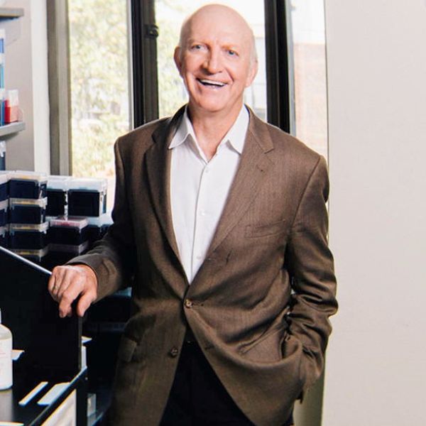
Meenhard Herlyn studies the normal and malignant tissue environment to develop rational approaches to cancer therapy, with a focus on melanoma, the most aggressive form of skin cancer.
Born and educated in Germany, Herlyn received his D.V.M. at the University of Veterinary Medicine, Hanover in 1970 and went on to receive a D.Sc. in medical microbiology at the University of Munich in 1976. He came to The Wistar Institute as an associate scientist in 1976, where he worked in the emerging field of monoclonal antibodies, a technology that formed the basis of a portion of today’s new targeted therapeutics. In 1981, Herlyn became an assistant professor and established a laboratory that is, today, one of the best-known research groups on the study of melanoma biology.
The Herlyn Laboratory

The Herlyn Laboratory
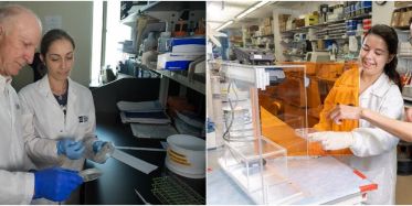
The Herlyn laboratory at The Wistar Institute focuses on the biology that underlies melanoma. Their efforts have pioneered the use of the three-dimensional “artificial skin” cultures to study the behavior of both tumor and normal cells that sustain tumor growth, a system known as the tumor microenvironment. The Herlyn Laboratory has transformed the scientific understanding of stem cells as they relate to cancer. Their work on the networks of signaling pathways in melanoma has formed the basis of numerous approved therapies.
-
Staff Scientists
Haiyin Li, Ph.D.
Haiwei Mou, Ph.D.
Eric Ramirez-Salazar, D.Sc. -
Postdoctoral Fellows
Gatha Thacker, Ph.D.
Qiuxian Zheng, Ph.D. -
Predoctoral Trainee
Jessica Kaster
-
Bioinformatics Technician
Yeqing Chen, M.S.
-
PDX Core Manager
Monzy Thomas, Ph.D.
-
Wistar Research Assistants
Ling Li, M.D.
Min Xiao, M.S. -
Research Assistants
Finley Medina
Veronika Yakovishina
Maggie Dunne -
Undergraduate Students (UPenn)
Hannah Hamdani
Vincent Ni
Elaine Tanel -
Technician
Abdiel Mandella Reynolds
-
Lab Coordinator
Jessica Kohn
Resources
CELL LINES
Due to the high demand for our cell lines, the Herlyn lab is pleased to collaborate with Rockland Immunochemicals Inc. for distribution.
Please contact Rockland customer service directly about acquiring our melanoma cell lines.
For help selecting lines, see the mutational chart below or search using Rockland’s website. For any questions, concerns, help, or other issues, please email Min Xiao at Wistar or contact customer service at Rockland.
STR PROFILES
Cell authentication has received considerable attention recently as more and more reports of cell line cross contaminations and misidentifications have come to light. As such, in 2008, we implemented and routinely perform Short tandem repeat (STR) profiling using AmpFlSTR® Identifiler® PCR Amplification Kit (Catalog Number 4322288) by Life Technologies which uses loci consistent with all major worldwide STR standards. PCR amplification and STR allele separation and sizing is performed by the Wistar Genomics Facility. Profile interpretation is performed in the Herlyn lab by interrogating the resulting DNA fingerprint to our internal database which includes over 1000 fingerprints, primarily Wistar Melanoma but also commonly used cell lines such as HeLa and 293T cells. The STR profile is provided here as a reference comparison to your results.
For additional inquiries, please email Min Xiao.
-
Staff Scientists
Haiwei Mou, Ph.D.
Eric Ramirez-Salazar, D.Sc.
Gatha Thacker, Ph.D. -
Bioinformatics Research Assistant
Yeqing Chen, M.S.
-
Wistar Research Assistants
Ling Li, M.D.
Min Xiao, M.S. -
Research Assistants
Maggie Dunne, B.A.
-
Undergraduate Students (UPenn)
Vincent Ni
PDX
The laboratory has generated more than 600 patient-derived xenografts (PDX), which have been extensively characterized at the DNA, RNA and protein levels. A selection of the PDX is available through Envigo.
ADDITIONAL RESOURCES
For inquiries regarding any of the following techniques or resources, please email Meenhard Herlyn to be put in touch with the appropriate lab representative.
Research
The Herlyn Laboratory seeks to further define the various signaling pathways that work in cancer cells to discover new opportunities to inhibit cancer growth through targeted therapeutics. Since therapy is increasingly guided by the genetic aberrations in tumors, Herlyn and his colleagues are developing combinations of compounds that take into account the genetic signature of tumors, with the specific goal of individualized cancer therapy. Currently, the Herlyn Laboratory collaborates with pharmaceutical companies as well as academic chemists and structural biologists to select and further develop compounds for tumor inhibition. Tumor heterogeneity, i.e., the differences between cells within one tumor, among different tumor lesions of the same patient, or between patients even if the tumors are of similar genetic signatures, provides major challenges for future therapy. The laboratory is developing biological signatures of melanoma cells that take into account the various forms of heterogeneity.
Another major effort of the Herlyn Laboratory is the study of therapy resistance and tumor dormancy. Tumor cells can become dormant in primary tumors or at any time after metastatic dissemination and can persist in the dormant state for many years, allowing tumors to resist treatment. Herlyn’s working hypothesis is that defined tumor subpopulations are central to dormancy and drug resistance due to their slow turnover and their non-responsiveness to growth signals. His efforts seek to define how tumor cells escape dormancy for growth, invasion, and metastasis, and how to best develop strategies for therapy.
MODELING THE NORMAL AND DISEASED HUMAN TISSUE MICROENVIRONMENT
The Herlyn lab is differentiating multi-potent stem cells from the human dermis and reprogrammed stem cells into melanocytes to test the hypothesis that melanocyte stem cells are more prone to transformation than fully differentiated cells, and that neighboring cells and matrix in the microenvironment play critical roles in differentiation and transformation. The lab has developed a complex, three-dimensional model that mimics human skin, and are using it to reconstruct each step in the melanoma development and progression cascade. Genes associated with melanoma are overexpressed or silenced with shRNA constructs in lentiviral vectors and the lab increasingly uses cDNA and CRISPR libraries for our experiments. Ultraviolet light irradiation is mimicking the DNA damaging effect of sunlight. Skin reconstructs can also be grafted onto immunodeficient mice for long-term observation. Besides isolating melanocytes and keratinocytes from skin, we differentiate them from ‘induced pluripotent stem’ (iPS). This source also allows the lab to generate an intact human inflammatory and immune system in vivo, including from melanoma patients where we have cell lines or patient-derived xenografts (PDX). Studies on interactions among tumor cells, fibroblasts and endothelial cells are also done in 3-D models, in which cells are embedded into collagen to mimic the tumor microenvironment. Growing cells in organ-like models (organoids) induces major changes in gene expression similar to those in animals and patients, making such models superbly suited for studies of cell-cell signaling, matrix formation, and drug resistance.
THERAPEUTIC TARGETING OF SIGNALING PATHWAYS IN CANCER
The Herlyn lab is defining signal transduction pathways that are constitutively activated in melanoma and squamous cell cancer cells through autocrine and paracrine growth factors and genetic alterations. With shRNA and CRISPR/Cas9 in lentiviral vectors, the lab is identifying genes in tumor cells, stromal fibroblasts, and endothelial cells that are potential targets for therapy. In melanoma, the MAPK and PI3K pathways are primary targets for therapy, but other pathways are also explored for inhibition by small molecule compounds. Since therapy is increasingly guided by genetic aberrations in tumors, the lab is developing combinations of compounds that take into account the genetic abnormalities of tumors, with the long-term goal of individualized cancer therapy. In recent years, the lab has actively collaborated with pharmaceutical companies to obtain compounds in early stages of preclinical and clinical development. Increasingly, the lab is collaborating with academic chemists and structural biologists to select and further develop compounds for tumor inhibition.
TUMOR DORMANCY AND THERAPY RESISTANCE
Tumor cells can become dormant in primary tumors or at any time after metastatic dissemination and can persist in the dormant state for many years, allowing them to resist treatment. The working hypothesis of the Herlyn lab is that tumor-maintenance cells (tumor stem cells) are central to dormancy due to their non-proliferation or very slow turnover and their non-responsiveness to growth signals. The lab is delineating tumor dormancy in melanoma and characterizing subpopulations of cells with a major focus on slow-proliferating cells that have high proliferation potential hypothesizing that these cells are critical for dormancy and therapy resistance. The lab will then define how tumor cells escape dormancy for growth, invasion, and metastasis, and developing strategies for therapy. Using the lab’s unique 3-D melanoma, Herlyn and his lab determine how microenvironmental cues from the matrix or other cells such as B cells, macrophages, and endothelial cells drive gene activation, leading to a signaling cascade for proliferation and invasion. These studies will lead to in-depth investigations of tumor heterogeneity and the dynamic regulation of genes that define subpopulations with specialized biologic functions. The long-term goal is to develop strategies for two therapies, one for eliminating the bulk of the tumor, the other for small subpopulations that escape all major therapeutic strategies. Such combinations should achieve elimination of all tumor cells, which is required in melanoma because single tumor cells are capable of tumor induction in immunodeficient animals.
STEM CELLS AND MELANOMA
Multipotent stem cells with neural crest-like properties have been identified by our lab and others in the dermis of human skin. The stem cells display self-renewal capacity and differentiate into neural crest derivatives including epidermal pigment-producing melanocytes. Neural crest-like stem cells (NCLSC) share many properties with aggressive melanoma cells, such as high migratory capabilities and expression of neural crest markers. However, little is known about which intrinsic or extrinsic signals determine proliferation or differentiation of stem cells. In our studies we have focused on major developmental pathways. Notch signaling is highly activated in stem cells, similar to cells within melanoma spheres. Inhibition of Notch signaling reduces proliferation of stem cells, induces cell death, and down-regulates non-canonical Wnt5a, suggesting that the Notch pathway contributes to maintenance and motility of the stem cells. In 3-D skin reconstructs, canonical Wnt signaling promotes differentiation of stem cells into melanocytes. This differentiation is triggered by the endogenous Notch inhibitor Numb, which is upregulated in the stem cells by Wnt7a derived from UV-irradiated keratinocytes. These studies reveal a crosstalk between the two conserved developmental pathways in human skin and highlight the role of the skin microenvironment in driving the generation of stem cells, and possibly tumor-initiating cells. They also provide a rationale for identifying novel targets for therapy among those groups of genes that are intimately involved in melanocyte development and highly expressed in melanoma while being largely absent in normal melanocytes.
Collaborations
The Herlyn laboratory has a long history of collaborations with members of the Penn/Wistar campus, particularly those who have had an interest in melanoma. Additionally, the lab has partnered with several outreach organizations around the world to support melanoma research. Learn more about these collaborations.
Funding
The Herlyn laboratory is supported by a variety of grants to study melanoma.
Contact Us
The Herlyn laboratory can provide access to several additional resources. To request a copy of a research paper published by the Herlyn laboratory, or for additional lab resources, contact:
Meenhard Herlyn
215-898-3950
The Wistar Institute
3601 Spruce Street
Philadelphia, PA 19104
Herlyn Lab in the News
Selected Publications
Dasatinib resensitizes MAPK inhibitor efficacy in standard-of-care relapsed melanomas
Rebecca VW, Xiao M, Kossenkov A, Godok T, Brown GS, Fingerman D, Alicea GM, Wei M, Ji H, Portuallo, M, Bravo J, Elad, V, Chen Y, Toska, E, Fane ME, Couts, KL, Villanueva J, Nathanson K, Arun, KM, Warrier, G, Yin, X, Ding, J, Liu Q, Goyal, Y, Gopal YNV, Davies MA, Herlyn M. Dasatinib resensitizes MAPK inhibitor efficacy in standard-of-care relapsed melanomas. iScience, (2026).
DR5 CAR-T cells target melanoma and suppress MDSCs with minimal toxicity. Molecular Therapy
Wang, H, Liu, S, Yang, J, Zhang, X, Goncalves, B, Zhen, Q, Mao, H, Yang, J, Guo, Y, Huang, L, Scholler, J, Liu, F, Miao, F, Zhang, C, Karakousis, G, Huang AC, Mitchell T, Amaravadi, R, Schuchter L, Fan, Y, Milone, M, Guo, W, June C, Herlyn, M, Xu, X. DR5 CAR-T cells target melanoma and suppress MDSCs with minimal toxicity. Molecular Therapy, (2026).
Spatiotemporal Profiling Defines Persistence and Resistance Dynamics during Targeted Treatment of Melanoma
Rubinstein JC, Domanskyi S, Sheridan TB, Sanderson B, Park S, Kaster J, Li H, Anczukow O, Herlyn M, Chuang JH. Spatiotemporal Profiling Defines Persistence and Resistance Dynamics during Targeted Treatment of Melanoma. Cancer Res. 2025 Mar 3;85(5):987-1002. doi: 10.1158/0008-5472.CAN-24-0690. PMID: 39700408


