Tag: Auslander

Wistar’s Dr. Noam Auslander Awarded $600K V Foundation Grant to Identify Connections between Gut Microbial Genes & Melanoma Immunotherapy

PRESS RELEASE
Dr. Auslander’s project will investigate the effects of microbial proteins on immunotherapy responses

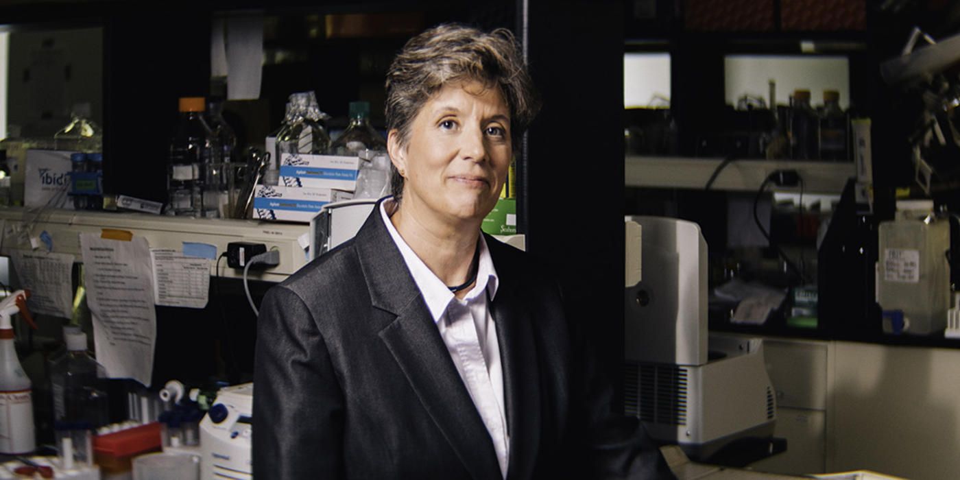
PHILADELPHIA — (Thursday, June 27, 2024) — The Wistar Institute’s Noam Auslander, Ph.D., assistant professor in the Molecular and Cellular Oncogenesis Program at the Ellen and Ronald Caplan Cancer Center, was awarded a $600,000 Women Scientists Innovation Award for Cancer Research grant from the V Foundation for Cancer Research to support the next three years of her research, which will use proteins of gut bacteria to predict immunotherapy benefit in melanoma. Dr. Auslander plans to analyze microbial proteins from the guts of patients to determine how they may drive melanoma immune responses, with the ultimate goal of improving the clinical benefits of immunotherapy.

“The V Foundation plays a crucial role in supporting and enabling transformative cancer research,” said Noam Auslander. “This prestigious award allows us to use computational biology methods to gain insights from complex data sets that can allow clinically impactful discoveries. Our goal through this project is to determine how patterns in the gut microbiome of melanoma patients determine immunotherapy responses. The V Foundation’s philanthropic support allows us to dig deep into this question using different computational methods, and, with the support from our clinician and experimental collaborators, potentially improve future clinical decisions and treatment outcomes.”

The V Foundation for Cancer Research was founded in 1993 by the late Jim Valvano, ESPN broadcaster and renowned basketball coach, and has allocated more than $353 million in grants for cancer research across the nation. By supporting the most promising cancer research projects from exceptional scientists of diverse backgrounds, The V Foundation has empowered investigations into cancer types of all stages — from the lab to the clinic.

“The V Foundation is honored to support Dr. Auslander’s innovative cancer research addressing a major unmet need in predicting responses to melanoma treatment. We look forward to seeing the novel findings her work will uncover and the impact it will have on patients in the future,” said Kara Coleman, Ph.D., Vice President of Research and Grants Administration at the V Foundation.

Dr. Auslander’s high-impact data research in melanoma immunotherapy happened due to generous V Foundation support.

“Dr. Auslander’s research identifies valuable patterns in human biology through large-scale data analysis with advanced computational techniques,” said Dario C. Altieri, M.D., Wistar president and CEO, director of the Ellen and Ronald Caplan Cancer Center, and the Robert and Penny Fox Distinguished Professor. “By investing in the burgeoning field of machine learning, this grant from the V Foundation accelerates the use of machine learning in transformative science.”

For a printer-friendly version of this release, please click here.

ABOUT THE WISTAR INSTITUTE:

The Wistar Institute is the nation’s first independent nonprofit institution devoted exclusively to foundational biomedical research and training. Since 1972, the Institute has held National Cancer Institute (NCI)-designated Cancer Center status. Through a culture and commitment to biomedical collaboration and innovation, Wistar science leads to breakthrough early-stage discoveries and life science sector start-ups. Wistar scientists are dedicated to solving some of the world’s most challenging problems in the field of cancer and immunology, advancing human health through early-stage discovery and training the next generation of biomedical researchers. wistar.org
Continue reading

Wistar Melanoma Researchers Discuss Risks and Solutions for Melanoma Awareness Month

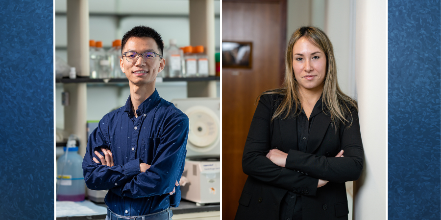
Three of The Wistar Institute’s foremost melanoma researchers: professor Meenhard Herlyn, D.V.M., D.Sc.; associate professor Jessie Villanueva, Ph.D.; and assistant professor Noam Auslander, Ph.D. discussed the progress and potential in melanoma research. Each brings their own distinct expertise to the field of melanoma research with decades of combined experience, and in reflecting on the state of the field, Drs. Herlyn, Villanueva, and Auslander covered both how they came to melanoma research and how they continue to tackle the challenge of this disease every single day at Wistar.

There are a lot of cancers out there. What brought you to melanoma?

Dr. Noam Auslander: As someone who works on the computational side of things, I was attracted to melanoma research mainly because of the quantity of data. In science generally but in computational science in particular, more data is better — because that allows researchers to design high-fidelity models, which, with cancer, can lead to all sorts of benefits, like predictions of who will respond to what therapy, or which genetic patterns are implicated in a cancer.

I can access and analyze melanoma data in large batches simply because there’s a lot of it. Part of that is because it’s a common cancer — which isn’t a good thing — but because it’s both common and a subject of study for more than 40 years, that allows my team and I to improve our models.

Dr. Jessie Villanueva: For me, melanoma research began as pure scientific interest. Melanoma is an aggressive cancer, and when I started as a postdoctoral fellow, there were no approved targeted therapies or immunotherapies; if chemotherapy, radiation, and surgery all failed, there really weren’t other options.

That problem attracted me to the field as a scientist who wants to solve problems, and shortly afterward, the professional interest became a personal one: a childhood friend whom I’d known since kindergarten was diagnosed with melanoma, and not long after that, so was my uncle. Unfortunately, my uncle passed away, but my friend survived, and that combination of loss and hope solidified melanoma as something I wanted to dedicate myself toward working against.

Dr. Meenhard Herlyn: My story is not so inspiring. I was young — so I suppose it was something like a hundred years ago — but my boss told me to help him with a melanoma project, and that was that. But I was very lucky, because that project involved a man named Wallace Clark: a great pathologist of the disease, whose research laid the foundation for much of what we know today about melanoma. Much of his work was characterizing these melanoma cells under a microscope — a necessary first step — and thinking of stories in his mind about how they might behave. Characterizing and theorizing. So as a young scientist, I thought to myself, “we must find a way to fill in these stories with real data.” And I’ve followed that ever since.

There are other skin cancers; melanoma is just a subtype. What makes it so dangerous?

J.V.: Melanoma comes from cells that originally have an innate level of pluripotency (the ability to transform into different cell types); they have remarkable migratory abilities; and they give rise to a diverse array of cell types throughout the body. When those cells become cancerous, they are highly plastic and skilled at adapting to their environment. This plasticity also allows melanoma to evade treatment and become drug-resistant. Drug resistance is a big problem in the field; often when using drugs targeting one pathway, the tumors find an alternative pathway to exploit.

By collectively studying all the inner workings of melanoma — like its genetics (the kind of mutations it collects), epigenetics (how genes are turned on or off), and signaling pathways (controlling processes like cell growth, proliferation, and survival) — we aim to develop strategies that prevent tumors from evading treatment. We’ve made great progress treating melanoma, but tumors still develop strategies to bypass therapies. This ongoing challenge drives our relentless search for innovative and effective solutions, fueled by the hope of achieving cures and improving the lives of melanoma patients.

N.A.: Melanoma is associated with an unusually high inter- and intra-tumor heterogeneity; the mutational profile is exceptionally complex between different melanoma cells and even within melanoma cells. That’s why large-scale data analysis of melanoma with computational models isn’t just important but necessary — patterns that can help us fight this cancer exist, but distinguishing between patterns and noise both within a tumor and between tumors requires the help of advanced computational techniques.

Meenhard has talked about how we need to listen to cells, and that’s how I try to help Meenhard & Jessie’s work: by fine-tuning computer systems to listen for signals amid the chaos in cancer.

M.H.: We also have to remember that the cells that become melanoma are highly mobile by their very nature. As Jessie said, melanocytes have a certain amount of innate plasticity, which contributes to the cancer’s aggression once a melanocyte goes from normal to cancerous.

But that wouldn’t necessarily be as big a problem if it weren’t for these cells’ motility. When you have aggressive cancer cells moving throughout the body, that creates a situation that lends itself to metastasis. A skin cancer that isn’t melanoma doesn’t present as much danger because it’s probably more localized; I’m not saying that’s not serious, but a non-metastatic tumor on the skin is a lot easier to treat — at the simplest level, you just cut it off. With melanoma, once that diagnosis comes, the clock is ticking to stop the cancer before the metastatic impulse gets out of control.

More people are getting melanoma, with U.S. incidence up by more than 50% since 1999. Why do you think that is, and how can people protect themselves?

J.V.: The short answer is that we don’t yet know for sure — there are several ongoing epidemiological studies which we expect will provide clear answers. Lifestyle is a big part of it. Outdoor activity can be healthy; however, being outdoors means more sun & UV exposure. Anecdotally, since the pandemic, we’ve noticed more people spending more time outdoors. And that’s a risk factor.

We’re seeing a sharp increase in melanoma for young people, particularly young women. Cancer tends to be correlated with age — the older we get, the higher the probability of having cancer — but melanoma is the most frequently developed cancer in people in their 20s and 30s.

M.H.: I agree that lifestyle is probably a big factor in the increase in cases. Everything from tanning beds to taking a vacation to lie on the beach is going to give UV rays more opportunity to cause damage that could lead to melanoma. Sunlight feels good to everyone, but unprotected exposure is harmful. People get addicted to damaging UV because our skin secretes endorphins when exposed to UV, and that’s more reason to be cautious.

It’s true that people with less melanin in their skin are more at risk — which is why, for example, more leisure travel from countries in the Global North to equatorial regions that get more sun probably causes more melanoma overall — but everyone has skin, which means anyone can get melanoma. And that’s why awareness of exposure risk is so important.

Revealing Biology’s Hidden Patterns: Wistar’s Dr. Noam Auslander on the Power and Potential of Machine Learning

Dr. Noam Auslander, Ph.D., is assistant professor of the Molecular and Cellular Oncogenesis Program at the Ellen and Ronald Caplan Cancer Center. She focuses on developing machine learning methods to understand the factors driving cancer development and to identify patterns that can improve cancer diagnosis and treatment.

“If you define your problem correctly, and you have enough data, you have the ability to learn something very complex that you cannot see with your eyes.”

How would you explain the difference between artificial intelligence and machine learning to somebody who is not a scientist?

Artificial intelligence is more general term. Any software that imitates the human learning system is artificial intelligence. If you build a robot, and that robot does nothing but respond to your requests, that’s artificial intelligence. Machine learning is a field of study contained within artificial intelligence that involves creating sets of algorithms that can be used to learn a particular task, independent of receiving instructions from humans.

As your field has advanced, how much of that advancement has been a matter of increased computing power versus improved methods?

It’s both of those things combined. Increased computing power has allowed algorithms created 15 or 20 years ago to suddenly become very efficient, very good. These older neural networks had architectures that consumed too much computing power at the time, but once we had the GPUs, they started to work much better. And then based on that there has been an explosion of new research. The algorithms have evolved even more, making them much, much better.

What role do you see for machine-learning models in biomedical data analysis and research?

Our models can extract more information and identify more patterns in data than humans could on their own. Right now, people are building models that will do things like predict clinical outcomes, predict biological factors, and understand more about biology. I think that’s very promising, because if you define your problem correctly, and you have enough data, you have the ability to learn something very complex that you cannot see with your eyes. But still, it requires a person who understands the data, understands what they are doing, and understands how to use the model correctly.

How do you develop models that can be used to generate meaningful insights about real-world data?

We first need to understand the question or problem we’re trying to address, and we need to understand the data well enough to represent it correctly in the algorithm. This usually means talking with the clinicians or the biologists to understand what they’re trying to do. We also need to understand how we define a good performance. Is the goal to build a test that can be used in the lab or in the clinic? Or are we trying to learn something new in biology? All of these factors go into designing the model.

What makes some data sets better suited to a machine learning approach than others?

In general, the more data we have, the more amenable it is for these methods, especially if it’s good, clean data. But there are also scenarios where you can take a model that’s been trained for one thing and apply it to another task. A good example is imaging data, like radiology. You can take a pre-trained model for imaging that has already looked at a lot of data. And instead of training the entire architecture, you can train a part of it to only recognize the specific thing you are trying to recognize. You’re using technology that has already learned from other problems that you had much more data for, and this makes it much, much easier.

What’s your biggest frustration you encounter when developing and training models?

It’s almost always not enough data. That can lead to overfitting, which means the model stays too close to the training data set and can’t begin to generalize and make the predictions that allow it to work independently. Or sometimes the data is too complex, we can’t trust it, it’s not annotated correctly, or there are clinical variables that are notated differently by different clinicians. Those kinds of things make it very difficult for us.

How do you keep up with all the changes in your field?

The area of machine learning is moving very fast, so we have to keep track of a lot of literature and a lot of new technology. It’s impossible to follow everything that happened even in the last year — if you’re two to five years behind, that’s pretty good. At the same time, it’s a very interdisciplinary field, so for every project we do, we have to keep up with the research in at least two different disciplines. So, in a way, we are keeping up with at least twice as much as what normal researchers do.

What do you think is the most fun or interesting thing about what you do?

It’s always fun and interesting to work in an area that’s changing so fast — you can be the first to do a lot of things. If you think of an important problem or question, you can be the person to address it. And because there is so much data being generated, we can make real biological discoveries, find out completely new things, without relying on a lab. We can use data that’s already out there and find out something that’s completely new.

The type of work you do requires a lot of creativity and problem solving. When you feel stuck on a problem, how do you get your creativity flowing again to look at the problem in a new way?

When I get stuck on a problem, like part of an algorithm not working, I leave it for a while. I’m a runner, so sometimes I’ll go for a run, and while I’m running I’ll have better ideas come to me. I think it’s always good to stop looking at the problem. Leave it for a while, then come back and take a fresh look.

For more information, email comm-marketing@wistar.org

Wistar Scientists Identify Esophageal Cancer Biomarkers

Dr. Noam Auslander and authors trained a neural network to identify cancer risk from microbes.

PHILADELPHIA—(Dec. 5, 2023)—Wistar scientists have developed a new tool that can help identify cancer-associated microbes by using machine learning technology. Under the leadership of Dr. Noam Auslander — assistant professor in the Ellen and Ronald Caplan Cancer Center’s Molecular & Cellular Oncogenesis Program — the group has analyzed short read RNA-sequencing data to detect biomarkers for esophageal carcinoma, or ESCA. Their paper, “Microbial gene expression analysis of healthy and cancerous esophagus uncovers bacterial biomarkers of clinical outcomes,” was published in International Society for Microbial Ecology Communications.

Tumor microenvironments are often analyzed using RNA sequencing, or RNAseq, which identifies mRNA in a population of cells to find which genes are being expressed. Theoretically, RNAseq data can reveal the expression of microbial genes in cancerous tissue, which could help to identify microbiome shifts that might be playing a role in the cancer’s development. But RNAseq “reads” — the physical lengths of genetic data that correspond with gene expression — are often quite short, posing a challenge for classifying them into diverse microbial genetic origins. Assembling the short RNAseq reads into longer contiguous segments that can be associated with a vast array of potential origins — be they human, viral, or bacterial — to identify specific microbes whose expression correlates with ESCA is computationally challenging.

That’s where Dr. Auslander and her group decided to intervene by training a convolutional neural network, a type of machine-learning technology that can be taught to train itself to accurately assess large quantities of data. The team, using large publicly available datasets of characterized short-read data, trained the network to sort short-read RNAseq data by its likely origin: human, viral, or bacterial. Their model sought to pare down the number of short reads that would need to be assembled for identification, which would reduce the computational load of screening for microbial influences in cancer tissue.

Once the model was trained, its sorting capabilities allowed the group to selectively analyze ESCA tissue for reads of microbial origin and compare those data with apparently healthy esophageal tissue. Auslander’s team found several instances of microbial expression present in ESCA with significantly less incidence in apparently healthy esophageal tissue.

In particular, they found that nearly half of the microbial genes over-expressed in cancer originated from bacteriophages, which are viruses that infect bacteria; this finding may indicate that viruses infecting microorganisms within the tumor microenvironment facilitate ongoing cancerous gene expression.

The team also identified patient outcome predictors amid the data. Bacterial iron-sulfur proteins were found to impact human genes involved in ferroptosis — a type of cell death pathway that’s modulated by iron — which predicted poor prognosis in ESCA patients. Microbial genes involved in mitochondrial reprogramming were also found to predict ESCA patient prognosis.

“By building on our previous work, our team has successfully leveraged machine learning to dive deeper into what’s going on inside cancer,” said Dr. Auslander. “While it’s always important to remember that correlation does not equal causation, the associations we’ve been able to find between certain microbial genes and ESCA will allow scientists to further understand the mechanics of esophageal cancer — which is the first step in stopping it.”

Co-authors: Daniel E. Schäffer of Carnegie Mellon University, The Wistar Institute, and the Massachusetts Institute of Technology; Wenrui Li of The University of Pennsylvania; Abdurrahman Elbasir and Dario C. Altieri of The Wistar Institute; Qi Long of The University of Pennsylvania; and Noam Auslander of The Wistar Institute.

Work supported by: National Cancer Institute grant numbers R00CA252025 and P30-CA016520 and National Institute on Aging grant number RF1-AG063481.

Publication information: “Microbial gene expression analysis of healthy and cancerous esophagus uncovers bacterial biomarkers of clinical outcomes,” published in International Society for Microbial Ecology Communications.

###

The Wistar Institute is the nation’s first independent nonprofit institution devoted exclusively to foundational biomedical research and training. Since 1972, the Institute has held National Cancer Institute (NCI)-designated Cancer Center status. Through a culture and commitment to biomedical collaboration and innovation, Wistar science leads to breakthrough early-stage discoveries and life science sector start-ups. Wistar scientists are dedicated to solving some of the world’s most challenging problems in the field of cancer and immunology, advancing human health through early-stage discovery and training the next generation of biomedical researchers. wistar.org

Wistar Scientists Discover Innate Tumor Suppression Mechanism

PHILADELPHIA — (MAY 4, 2023) — The p53 gene is one of the most important in the human genome: the only role of the p53 protein that this gene encodes is to sense when a tumor is forming and to kill it. While the gene was discovered more than four decades ago, researchers have so far been unsuccessful at determining exactly how it works. Now, in a recent study published in Cancer Discovery, a journal of the American Association for Cancer Research, researchers at The Wistar Institute have uncovered a key mechanism as to how p53 suppresses tumors. By using a genetic variant of p53 and comparing what that variant failed to accomplish with what the healthy “wild type” p53 gene could do, the researchers discovered the mechanism by which p53 triggers immune function that, in turn, kills the tumor.

“The paradigm shift is that, instead of asking ‘What does p53 do’ we were able to use a lesser-functioning but cancer-predisposing genetic variant in African Americans to tell us ‘What does p53 not do when it doesn’t suppress cancer?’” said Maureen E. Murphy, Ph.D., senior author on the paper and deputy director of the Ellen and Ronald Caplan Cancer Center and Ira Brind Professor and program leader of the Molecular & Cellular Oncogenesis Program at The Wistar Institute.

Four and a half million people in the United States possess inherited, or germline, mutations in p53, which increases their risk of cancer. A small subset of these individuals have a mutation that leads to Li Fraumeni Syndrome, which results in their developing multiple tumors every few years, starting in childhood. Others with different p53 mutations possess what are called hypomorphs: a gene variant having a similar but weaker effect than the corresponding normal, or wild-type, gene. These people also develop cancer, but theirs is less aggressive, and they develop it later in life.

Murphy and her team decided to learn how p53 suppresses tumors by exploring how one particular hypomorph fails to suppress them. The researchers chose an African-specific variant called Y107H due to the fact that African Americans have the largest cancer burden of any ethnic group in the world. Their first hypothesis was that they could use the hypomorph to find which “downstream” genes—which p53 would ordinarily turn on—are critical for suppressing tumors. Their second hypothesis was that they could then screen for drugs that would kill the hypomorph tumors: Murphy’s group was able to accomplish both goals.

The researchers began by using CRISPR engineering to make a mouse model of their African-specific hypomorph Y107H. As expected, the mice with Y107H developed many forms of cancer and, as with humans who possess this variant, they started developing cancer in “middle age” (i.e., after 12-14 months of an average two-year lifespan).

Next, the researchers created tumor cell lines with their Y107H hypomorph, as well as cell lines with a hypomorph found in Ashkenazi Jewish populations, called G334R. They then compared which genes were turned on by normal, or wild type, p53 (to suppress the tumor) but not turned on by the two hypomorphs (which failed to suppress the tumor). The gene that met these conditions was PADI4. To confirm, they checked ten other hypomorphs—none of those variants turned on PADI4, either.

“It’s as though this was the key p53 target gene that, every time you have a genetic variant that predisposes you to cancer, it cannot turn on this gene,” said Murphy. She added that it makes sense that PADI4 would be implicated, because this gene helps the immune system recognize tumors. It does this by modifying components of tumor proteins so that they become citrulline, which is a non-natural amino acid. When the immune system recognizes citrulline as a foreign body, it attacks.

“Essentially, when a tumor cell goes from one cell to two and it’s not supposed to, p53 is alarmed, it turns on PADI4, and PADI4 says, ‘Immune system, you better come get me,’” said Murphy.

The final stages of Murphy’s research went beyond foundational research and looked toward helping cancer patients. First, the researchers used Wistar’s Molecular Screening and Protein Expression facility to identify drugs that would be effective against tumors with the Y107H hypomorph while sparing tumors with wild-type p53. Then, they looked for a way to predict which patients would respond to immunotherapy and which would not. Ordinarily, in order to do this, they would need many more human tissue samples from African Americans than they had. So instead, they turned to machine learning.

“Enter Noam Auslander, Ph.D., who is a brilliant machine learning artificial intelligence person here at Wistar,” said Murphy. “She said, ‘Let me find the genes that p53 and PADI4 control together using bioinformatic approaches and create a gene signature.’”

To do this, Auslander analyzed 60,000 tumors in the TCGA database and identified five genes that were coregulated together by wild type p53 and PADI4 and that the Y107H hypomorph couldn’t turn on. Upon further analysis, she found that this five-gene signature predicted cancer survival, immune infiltration into the tumor, and who would respond to immunotherapy.

Murphy believes that identifying this gene signature through machine learning was what pushed her team’s paper from a scientific breakthrough to a medical game-changer. “We’ve not only said we have an important p53 target gene, but we also have an important five-gene signature that will actually tell us who will respond to immunotherapy and who won’t, and p53 is at the core of this signature.”

She also believes that this research could only have been performed at an institution like Wistar, because collaboration was so crucial. “If you look at the authors on this, I have immunologists who did the immunology; I have machine learning people who did the bioinformatics; and I have drug screening people who did the compound screens,” said Murphy.

“Wistar is just a thrilling place where everyone here is saying, ‘Here’s how I can help your research.’ It makes all the difference.”

Co-authors: Alexandra Indeglia, Jessica C. Leung, James F. Dougherty, Nicole Clarke, Nicole A. Kirven, Chunlei Shao, Thibaut Barnoud, David Y. Lu, Isabela Batista Oliva, Qin Liu, Joel Cassel, Noam Auslander, Cindy Lin, Tyler Yang, Daniel Claiborne, Yulia Nefedova, Toshitha Kannan, and Andrew V. Kossenkov from The Wistar Institute; Sven A. Miller, Lei Ke, and John Karanicolas from Fox Chase Cancer Center; Julia I-Ju Leu from the Perelman School of Medicine at the University of Pennsylvania; Scott Lovell and Lijun Liu from the Del Shankel Structural Biology Center at The University of Kansas; Kevin P. Battaile from the New York Structural Biology Center; and Peter Vogel from St. Jude Children’s Research Hospital.

Work supported by: National Health Institutes (NIH) grants CA102184 to M.M., CA238611 to M.M., R00CA241367 to T.B., and P30CA006927 to J.K.

Publication information: An African-Specific Variant of TP53 Reveals PADI4 as a Regulator of p53-Mediated Tumor Suppression, Cancer Discovery, 2023. Online publication.

###

The Wistar Institute, the first independent, nonprofit biomedical research institute in the United States, marshals the talents of an international team of outstanding scientists through a culture of biomedical collaboration and innovation. Wistar scientists are focused on solving some of the world’s most challenging and important problems in the field of cancer, infectious disease, and immunology. Wistar has been producing groundbreaking advances in world health for more than a century. Consistent with its legacy of leadership in biomedical research and a track record of life-saving contributions in immunology and cell biology, Wistar scientists’ early-stage discoveries shorten the path from bench to bedside. wistar.org.

Wistar Scientists Uses Artificial Intelligence to Identify Viruses Related to Cancer

Some cancers are linked to viral infections. Studying viruses found in tumor cells can reveal important information in the development of more effective cancer treatments. Wistar researchers developed a tool to study the expression of cancer-related viruses through artificial intelligence. In a recent paper published in Nature Communications by Noam Auslander, Ph.D., assistant professor, Molecular & Cellular Oncogenesis Program, Ellen and Ronald Caplan Cancer Center, and her lab, created the technology called viRNAtrap as an innovative method that identifies viruses from human RNA sequences and rapidly characterizes viruses expressed in tumors.

Wistar discussed viRNAtrap and its creation with Dr. Auslander to find out more about how this novel technology impacts research on cancer and other viral diseases.

Q: What inspired this research to develop a new platform analyzing viral expression linked to cancer? Is this a one-time study or part of a larger project?

A: I have always wanted to investigate viruses that cause cancer or correlate with cancer outcomes. As a trainee I worked in computational labs that studied cancer or viruses (but not both) and used different tools for these studies. In my lab I incorporate those tools, allowing the development of this framework. This is a major research direction in my lab, and we have follow-up projects that are looking into related questions.

Q: What is viRNAtrap? How did you and your team come up with this name?

A: My postdoc Dr. Abdurrahman Elbasir and I came up with the name. It combines vi- (for virus), RNA (for RNA sequences), and trap (because we “trap” viral RNA sequences that are difficult to identify).

Q: What can viRNAtrap do?

A: It’s a software to identify viruses from short RNA sequencing reads – taking small fragments of the genome then assembling longer sequences of viruses that are expressed in a tissue.

Q: What were your methods in creating this framework? Were there any challenges that arose during the process?

A: As a postdoc I worked on an AI software to identify viruses, but this platform was based on longer sequences coming from a different technology. The read length was and is a major bottleneck for viRNAtrap. Dr. Elbasir managed to train a deep learning model — that’s a model that is built using neural networks that can distinguish viral reads from human reads fairly well using reads as short as 48bp. This model and the proof of concept that it could be built were critical for this research. Based on this model, we built the viRNAtrap framework that identifies viral reads and assembles longer sequences (contigs) from which known and new viruses can be characterized.

Q: How did you verify viRNAtrap works?

A: The model was validated and tested with an independent test dataset. The whole framework was verified using cases with known cancer viruses in the TCGA. We also had an experimental validation for one of the new viruses that we found in ovarian cancer, through a collaboration with Dr. Rugang Zhang’s lab, who verified that this virus is expressed in cell lines.

Q: Was there anything surprising that viRNAtrap detected?

A: There were a couple of very surprising viruses viRNAtrap detected, including some plant and insect viruses that were found in tumor tissues. The most notable of which was an insect virus that we found in 25% of endometrial cancer samples. If this association is real and not due to some unidentified contamination of the TCGA samples, this could be a very important discovery.

Q: How can this tool be used in biomedical studies to help prevent/combat cancer and other diseases?

A: We all know that viruses are a major health concern, and that they contribute to many diseases. However, viruses are really difficult to study with current sequencing technologies as they evolve rapidly and accumulate many mutations. Using this tool, we can identify new viruses in disease tissues even if they are divergent and mutated. We can therefore find viruses that drive or modulate diseases, which can lead to new diagnosis, vaccination, and treatment strategies.

Dr. Noam Auslander Wins Michelson Prize

The annual award supports early career investigators and their work advancing in the immunology, vaccine, and immunotherapy space.

Noam Auslander, Ph.D., Assistant Professor in the Molecular & Cellular Oncogenesis Program of the Ellen and Ronald Caplan Cancer Center at The Wistar Institute, was named a Michelson Prize laureate by The Michelson Medical Research Foundation and the Human Immunome Project.

The Michelson Prize: Next Generation Grants are awarded annually to promising early-career investigators with research projects focused on human immunology, vaccine discovery, and immunotherapy research. The awardees are selected by a committee of leading scientific experts from around the world and receive $150,000 to support their innovative science. Auslander is one of four winners that make up the 5th cohort of Michelson Prize awardees.

Auslander and her lab are developing artificial intelligence approaches to detect microbes in cancer and immune diseases. This work provides important insight into immune responses of patients to disease and disease outcomes – ultimately impacting the creation of vaccines and immunotherapies. 

The Lab of the Future: AI’s Impact on Biomedical Research

The evolution of machine learning and artificial intelligence is changing the way the contemporary lab looks and functions. Researchers today are discovering that breakthroughs can happen not only at the bench, but on the desktop.

Noam Auslander, Ph.D., assistant professor in the Molecular & Cellular Oncogenesis Program of Wistar’s Ellen and Ronald Caplan Cancer Center, conducts her research at the intersection of computer science and biomedical science. The interdisciplinary nature of Wistar provides fertile ground for her innovative lab to flourish and tackle research questions from a multifaceted and collaborative perspective. She uses advanced techniques to investigate genetic factors underpinning cancer evolution and viruses to improve diagnostics and therapeutics. As a computational scientist, Auslander applies the power of advanced computational platforms – artificial intelligence (AI) and machine learning (ML) software – to very intricate and complex biomedical data.

Wistar had a conversation with Auslander to find out more about how computer science impacts biomedical research on cancer and viral diseases and the evolution of the next generation research lab.

Can you define artificial intelligence and machine learning?

These are fields in computer science that involve algorithms allowing learning from a set of examples. This can be for instance, learning to make decisions based on data, or to transform one data into another form.

What is unique about using computer science and AI approaches in biomedical research? How far along is the field in biomedical science?

There are some biomedicine areas where AI approaches are very advanced, such as for prediction of protein structure or radiology. However, in other domains, such as for drug identification or precision medicine, it is still in its infancy. Some reasons for these differences are (1) how much data is available to build AI models (2) how much effort is invested to address a particular research question and (3) how well defined the problem is in terms of data and goals.

Why is using advanced computational methods important to biomedical research? What types of knowledge and data can be gathered and analyzed using AI?

We have a huge amount of data that is available to us, and datasets are being generated every day. Within these, there is hidden information that can benefit biomedicine if uncovered, and the only way to do this realistically is by applying computational approaches and improving methodologies that can harness these datasets.

What do you anticipate for the future of computer science techniques in a research space? What does innovation look like in your field?

We need advancements in algorithms and implementations that will make the most use of continuous improvements in computing power.

Just as libraries have dramatically changed with the proliferation of computers and tablets – do you see the lab of the future morphing more into computers and software and fewer test tubes and high-end imaging equipment?

It is already happening – almost every graduate program today is teaching some basic coding skills, and many wet-lab biologists today hire computational staff. This trend will probably continue.

What is one thing you wish people knew about harnessing the power of artificial intelligence for research?

I wish people would be careful and acquire appropriate training before using such tools in their research.

What impact do you hope your work will have on cancer and infectious disease?

We are searching for unknown organisms that are associated with cancers and other diseases. I hope that we will find viruses and bacteria that play a role in these diseases and have not been discovered previously.

How is your day-to-day schedule structured?

I oversee the projects in my lab and some ongoing projects with collaborators. There is a lot of processing data and making sure things are done and defined correctly. For scientific literature, I need to keep track of many fields, including cancer research, viruses and infectious diseases, and new computational advancements ¬– and that’s not easy.

What is your favorite part of work and Wistar?

The best part in work is when we find something that no one found before. Wistar is great because people here are very supportive and collaborative, which allows us to move forward, validate new findings, and apply our methods to other domains.

Do you collaborate a lot and how does your lab work together with other Wistar labs?

We are very collaborative, and we have different types of collaborations. Trainees in my lab decide if they work on collaborative projects and to what extent, and this motivates the ‘how’ my lab works with others.

What advice do you have for those interested in pursuing a career like yours?

Start coding early in life and find programs that allow comprehensive training in computer science and biology.

Wistar Scientists Identify Key Biomarkers that Reliably Predict Response to Immune Checkpoint Inhibitor Therapy for Melanoma

New research finds biological processes that improve prediction of therapeutic performance and provide a framework to develop predictors for this aggressive skin cancer.

PHILADELPHIA — (SEPTEMBER 19, 2022) — Immune checkpoint inhibitor (ICI) therapy is a type of treatment for melanoma, the deadliest form of skin cancer, which blocks proteins on tumor or immune cells that prevent the immune system from killing cancer cells. While this treatment has shown some clinical success in patients with advanced stages of melanoma, its efficacy depends on reliable predictors of a patient’s response to the therapy. Currently, the only FDA approved biomarker for ICI melanoma treatment is the tumor mutation burden assay, ¬but the mechanisms linking it to ICI remain unclear. However, new research now provides evidence of novel, reliable biomarkers that predict therapy response using advanced computer technology.

In a paper published in Nature Communications, Noam Auslander, Ph.D., assistant professor in the Molecular & Cellular Oncogenesis Program of Wistar’s Ellen and Ronald Caplan Cancer Center, and Andrew Patterson, graduate student in the Auslander lab, identify novel predictors of ICI therapy for melanoma. In particular, mutations in the processes of leukocyte and T-cell proliferation regulation show potential as biomarkers with reliable and stable prediction of ICI therapy response across multiple different datasets of melanoma patients.

“This work aims to identify better and more biologically interpretable genomic predictors for immunotherapy responses,” notes Auslander. “We need better biomarkers to help select patients that are more likely to respond to ICI therapy and understand what factors can help to enhance responses and increase those numbers.”

Using machine learning and publicly available de-identified clinical data, researchers investigated why some melanoma patients responded to ICI therapy and others did not. Patterson, first author on the paper, details that their research process involved training machine learning models on a dataset to predict whether a patient responds to ICI therapy, and then confirming that the model was able to continually predict response or resistance to this treatment over multiple other datasets.

The team found that leukocyte and T-cell proliferation regulation processes have some mutated genes that contribute to ICI treatment response and resistance. This knowledge could be used to identify targets to enhance responses or mitigate resistance in patients with melanoma.

“We were able to better predict if a patient would respond to ICI therapy than the current clinical standard method as well as extract biological information that could help in further understanding the mechanisms behind ICI therapy response and resistance.” Patterson explains.

The scientists intend to continue this work with the goals of increasing prediction accuracy, further understanding biological mechanisms underpinning patient resistance or responsiveness to ICI therapy, and determining whether the processes distinguished in the paper can also serve as predictors of ICI treatment response for other cancer types.

Co-author: Andrew Patterson

Work supported by: The results shown here are in whole or part based upon data generated by the TCGA Research Network: https://www.cancer.gov/tcga. The research reported in this publication was supported in part by the National Cancer Institute of the National Institutes of Health under Awards R00 CA252025 and P50 CA174523.

Publication Information: Mutated Processes Predict Immune Checkpoint Inhibitor Therapy Benefit in Metastatic Melanoma. Nature Communications, 2022. Online publication.

###

The Wistar Institute marshals the talents of an international team of outstanding scientists through a highly-enabled culture of biomedical collaboration and innovation, to solve some of the world’s most challenging and important problems in the field of cancer, immunology, and infectious diseases, and produce groundbreaking advances in world health. Consistent with a pioneering legacy of leadership in not-for-profit biomedical research and a track record of life-saving contributions in immunology and cell biology, Wistar scientists pursue novel and courageous research paths to life science discovery, and to accelerate the impact of early-stage discoveries by shortening the path from bench to bedside.

Scientists Drive Innovation at Wistar’s Ellen and Ronald Caplan Cancer Center

Wistar continues to be a dynamic environment prepared to tackle biomedical challenges in a collaborative, innovative, and inclusive culture. Read more about our Ellen and Ronald Caplan Cancer Center commitment to scientific career development, a diverse research community, and how previously introduced recruits are settling in and advancing impactful science.

ENHANCING CAREERS AND EXPANDING DIVERSITY

Italo Tempera, Ph.D., newly appointed Associate Director for Cancer Research Career Enhancement, was a postdoctoral fellow at Wistar and returned as an associate professor in the Gene Expression and Regulation Program in 2020. His research focuses on epigenetic mechanisms behind Epstein-Barr Virus (EBV). He was recently named associate director for Cancer Research Career Enhancement.

Tempera considers the time he spent at Wistar to be formative. With its very collaborative introductory environment, Wistar is an “… opportunity for our students not only to learn about our science but to get in contact with scientists.”

Furthermore, he outlines what he would like to accomplish in his new role. “We’re outstanding scientists and we have excellent mentors. The opportunities for our trainees to do an internship with different departments is something we want to push forward, and we want to expand the Cancer Biology Ph.D. program that we have now with Saint Joseph’s University.”

He shares that Wistar gave him the opportunity to grow as a scientist and advance in his research career. “When someone asks what was one of the most important aspects of a scientist’s pre- or post-doctoral training, my goal is for the trainee to think back and reply that being at Wistar has made all the difference.”

Jessie Villanueva, Ph.D., newly appointed Associate Director for Diversity, Equity, and Inclusion, joined Wistar first as a postdoctoral fellow and then was appointed assistant professor in the Molecular and Cellular Oncogenesis Program. Her work aims to identify targets for therapy to treat melanoma.

“Diversity leads to innovation and scientific excellence. New discoveries and scientific breakthroughs often rely on collaborations, and diverse teams are more creative and resourceful,” she shares.

For her new role, Villanueva aims to lead and inspire everyone at Wistar to integrate inclusion, diversity, and equity into all facets of the Institute. “Our goal is to continue fostering an inclusive community where everyone can develop to their full potential while contributing to Wistar’s mission of scientific discoveries.” To accomplish this, she plans to work with leaders and stakeholders across the Institute to identify challenges and areas for
improvement and propose strategies to address them.

“Diversity supports Wistar’s mission,” Villanueva asserts. She elaborates that many of the Institute’s scientific breakthroughs are largely impactful for biomedical sciences and human health, and these discoveries rely on “… outstanding scientists, trainees and staff with diverse backgrounds and skills who support Wistar’s goals wholeheartedly.”

ADVANCING IMPACTFUL SCIENCE

Nan Zhang, Ph.D., Assistant Professor, Immunology, Microenvironment & Metastasis Program, joined Wistar in September 2021 as an assistant professor and currently researches how immune cells play a role in tumor growth in abdominal cancers.

“Studying disease was always one of my passions,” Zhang shares as he describes both a personal and professional draw to cancer research. He began his career studying the immune system — particularly macrophages, a special population of white blood cells that removes unwanted materials in the body like harmful microorganisms or dead cells.

Upon completion of his postdoctoral position, Zhang felt that cancer in the peritoneal space — the area of the body encompassing the abdomen and the organs within it — would be a great direction to pursue for his future career because of its unique complexity and how it’s less understood relative to other focus areas for cancer research. This is what he works on now at Wistar.

Immersed in the Institute’s world class techniques, resources, and renowned scientists, Zhang continues to push forward his research to tackle how to use specialized cells called macrophages to combat tumors as a checkpoint therapy for cancer. He is also investigating immunological questions about the microenvironment of the peritoneal space and how this knowledge can help inform therapeutics and treatment development.

He shares, “Wistar is competitive, and the support in the Institute for junior faculty is great. We have meetings every week and this is an environment I really wanted for my career and research.”

Noam Auslander, Ph.D., Assistant Professor, Molecular & Cellular Oncogenesis Program, joined Wistar in June 2021 as an assistant professor and conducts her research at the intersection of computer science and biological science. She uses machine learning to investigate genetic factors underpinning cancer evolution to improve diagnostics and therapeutics.

“I work on cancer and viruses. Both are complex and have high mutation rates. As a computational scientist, it’s very interesting because there are a lot of computational challenges that can be investigated,” Auslander comments.

She joined The Wistar Institute because of its reputation and expertise, particularly in researching both cancer and viruses. She shares her experience during her first year, “It’s a small institute with a lot of opportunities to collaborate. It’s a very good environment and people are very helpful and supportive.”

Simultaneous to establishing and expanding her lab group, Auslander is currently looking into improving clinical prognosis to cancer and other diseases by uncovering unknown infectious agents and therapeutic biomarkers. To accomplish this, her lab applies the power of advanced computational platforms to very intricate and complex biomedical data to make these predictors of treatment responses more biologically interpretable. She says, “My main focus at the moment is to train my growing lab and develop frameworks to identify new viruses and eventually new microbiomes in cancer.”