Author: The Wistar Institute

The Wistar Institute Awarded Second National Science Foundation Grant to Expand Award-winning STEM Training Program

PRESS RELEASE

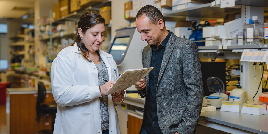
PHILADELPHIA — (May 14, 2024) — The Wistar Institute was awarded a $649,971 grant from the National Science Foundation (NSF) to support the continued expansion of its award-winning Biomedical Technician Training (BTT) Pre-apprenticeship Program. The grant supports Wistar’s reach to community colleges in New Jersey and Delaware who may have limited or no access to hands-on laboratory training and internships. This marks the second NSF grant supporting the Program’s continued expansion.

“As the Greater Philadelphia Region’s life science sector continues to expand, there is greater demand for laboratory technicians in both academic and industry labs,” said Dario Altieri, M.D., president and CEO, director of the Ellen and Ronald Caplan Cancer Center, and the Robert and Penny Fox Distinguished Professor of The Wistar Institute. “This 25-year program has offered career pathways to a candidate pool that is eager to join the life science sector and gain access to both entry-level and long-term careers here in Philadelphia.”

Created in 2000, The Wistar Institute’s BTT Program was initially designed as a two-summer, hands-on, mentored technician training program that prepared community-college students for positions in academic and biomedical, biotechnology, and pharmaceutical laboratories. Originally limited to students from Community College of Philadelphia (CCP), in 2021, Wistar received its first National Science Foundation (NSF) Advanced Technological Education (ATE) grant, Expansion, Curriculum Evolution, and Enhancement during BioTechnician Training (ExCEEd BTT), allowing it to expand from a cohort of 12 students from CCP to a cohort of 20 students from CCP and four other regional community colleges.

The latest grant, Tri-State ExCEEd BTT, will enable Wistar to bring its BTT Program to additional community colleges in New Jersey and Delaware starting in Summer 2025 through Summer 2027. The grant will expand the applicant pool in the Greater Philadelphia Region by including three additional community college collaborators, for a total of eight community colleges covering at least 10 counties in three states for Wistar’s BTT Pre-apprenticeship Program.

In ExCEEd BTT, students engage in a paid, accelerated, one-summer pre-apprenticeship training that includes a hands-on laboratory orientation at Wistar and two full-time, mentored experiences in academic and industry labs. Program graduates are prepared for immediate employment as laboratory technicians and may also continue training through Wistar’s registered Fox Biomedical Research Technician (BRT) Apprenticeship.

The BTT Program and BRT Apprenticeship provide training and research experiences not typically available to associate degree students, a segment of the workforce that is indispensable to support the success of an ever-expanding life science sector. Tri-State ExCEEd BTT supports Wistar’s commitment to building a diverse and inclusive life science sector talent pool. With this support from the NSF Wistar can expand its programming base and continue to train a diverse and underrepresented student population with limited access to life science research.

“By exposing students to Wistar science, we’re giving them access to the latest research, so they are ideally positioned for future careers,” explained Dr. Kristy Shuda McGuire, Dean of Biomedical Studies at The Wistar Institute. “Our approach is to work closely with faculty at community colleges to develop a cohesive program that offers students the foundational knowledge and the hands-on training they need to be successful. Then they put their knowledge and skills to work with two lab experiences with academic or industry collaborators here in the region doing cutting-edge science.”

Tri-State ExCEEd BTT will also add cell and gene therapy components to the current curriculum, provide a blueprint for incorporating a biotechnician pre-apprenticeship program into various biotechnology and science curricula at community colleges, and serve as a model for regional program expansion. New employer collaborators in two neighboring states will be recruited, allowing the registration of the apprenticeship for use nationally.

For a printer-friendly version of this release, please click here.

ABOUT THE WISTAR INSTITUTE:

The Wistar Institute is the nation’s first independent nonprofit institution devoted exclusively to foundational biomedical research and training. Since 1972, the Institute has held National Cancer Institute (NCI)-designated Cancer Center status. Through a culture and commitment to biomedical collaboration and innovation, Wistar science leads to breakthrough early-stage discoveries and life science sector start-ups. Wistar scientists are dedicated to solving some of the world’s most challenging problems in the field of cancer and immunology, advancing human health through early-stage discovery and training the next generation of biomedical researchers. wistar.org.

Continue reading

Revealing Biology’s Hidden Patterns: Wistar’s Dr. Noam Auslander on the Power and Potential of Machine Learning

Dr. Noam Auslander, Ph.D., is assistant professor of the Molecular and Cellular Oncogenesis Program at the Ellen and Ronald Caplan Cancer Center. She focuses on developing machine learning methods to understand the factors driving cancer development and to identify patterns that can improve cancer diagnosis and treatment.

“If you define your problem correctly, and you have enough data, you have the ability to learn something very complex that you cannot see with your eyes.”

How would you explain the difference between artificial intelligence and machine learning to somebody who is not a scientist?

Artificial intelligence is more general term. Any software that imitates the human learning system is artificial intelligence. If you build a robot, and that robot does nothing but respond to your requests, that’s artificial intelligence. Machine learning is a field of study contained within artificial intelligence that involves creating sets of algorithms that can be used to learn a particular task, independent of receiving instructions from humans.

As your field has advanced, how much of that advancement has been a matter of increased computing power versus improved methods?

It’s both of those things combined. Increased computing power has allowed algorithms created 15 or 20 years ago to suddenly become very efficient, very good. These older neural networks had architectures that consumed too much computing power at the time, but once we had the GPUs, they started to work much better. And then based on that there has been an explosion of new research. The algorithms have evolved even more, making them much, much better.

What role do you see for machine-learning models in biomedical data analysis and research?

Our models can extract more information and identify more patterns in data than humans could on their own. Right now, people are building models that will do things like predict clinical outcomes, predict biological factors, and understand more about biology. I think that’s very promising, because if you define your problem correctly, and you have enough data, you have the ability to learn something very complex that you cannot see with your eyes. But still, it requires a person who understands the data, understands what they are doing, and understands how to use the model correctly.

How do you develop models that can be used to generate meaningful insights about real-world data?

We first need to understand the question or problem we’re trying to address, and we need to understand the data well enough to represent it correctly in the algorithm. This usually means talking with the clinicians or the biologists to understand what they’re trying to do. We also need to understand how we define a good performance. Is the goal to build a test that can be used in the lab or in the clinic? Or are we trying to learn something new in biology? All of these factors go into designing the model.

What makes some data sets better suited to a machine learning approach than others?

In general, the more data we have, the more amenable it is for these methods, especially if it’s good, clean data. But there are also scenarios where you can take a model that’s been trained for one thing and apply it to another task. A good example is imaging data, like radiology. You can take a pre-trained model for imaging that has already looked at a lot of data. And instead of training the entire architecture, you can train a part of it to only recognize the specific thing you are trying to recognize. You’re using technology that has already learned from other problems that you had much more data for, and this makes it much, much easier.

What’s your biggest frustration you encounter when developing and training models?

It’s almost always not enough data. That can lead to overfitting, which means the model stays too close to the training data set and can’t begin to generalize and make the predictions that allow it to work independently. Or sometimes the data is too complex, we can’t trust it, it’s not annotated correctly, or there are clinical variables that are notated differently by different clinicians. Those kinds of things make it very difficult for us.

How do you keep up with all the changes in your field?

The area of machine learning is moving very fast, so we have to keep track of a lot of literature and a lot of new technology. It’s impossible to follow everything that happened even in the last year — if you’re two to five years behind, that’s pretty good. At the same time, it’s a very interdisciplinary field, so for every project we do, we have to keep up with the research in at least two different disciplines. So, in a way, we are keeping up with at least twice as much as what normal researchers do.

What do you think is the most fun or interesting thing about what you do?

It’s always fun and interesting to work in an area that’s changing so fast — you can be the first to do a lot of things. If you think of an important problem or question, you can be the person to address it. And because there is so much data being generated, we can make real biological discoveries, find out completely new things, without relying on a lab. We can use data that’s already out there and find out something that’s completely new.

The type of work you do requires a lot of creativity and problem solving. When you feel stuck on a problem, how do you get your creativity flowing again to look at the problem in a new way?

When I get stuck on a problem, like part of an algorithm not working, I leave it for a while. I’m a runner, so sometimes I’ll go for a run, and while I’m running I’ll have better ideas come to me. I think it’s always good to stop looking at the problem. Leave it for a while, then come back and take a fresh look.

For more information, email comm-marketing@wistar.org

Wistar Scientists Discover New Immunosuppressive Mechanism in Brain Cancer

PRESS RELEASE
Wistar’s Veglia lab identified how glioblastoma evades the immune system by inducing pro-tumor macrophages via a glucose based epigenetic modification.

PHILADELPHIA — (May 3, 2024) — The Wistar Institute assistant professor Filippo Veglia, Ph.D., and team, have discovered a key mechanism of how glioblastoma — a serious and often fatal brain cancer — suppresses the immune system so that the tumor can grow unimpeded by the body’s defenses. The lab’s discovery was published in the paper, “Glucose-driven histone lactylation promotes the immunosuppressive activity of monocyte-derived macrophages in glioblastoma,” in the journal Immunity.

“Our study shows that the cellular mechanisms of cancer’s self-preservation, when sufficiently understood, can be used against the disease very effectively,” said Dr. Veglia. “I look forward to future research on metabolism-driven mechanisms of immunosuppression in glioblastoma, and I’m hopeful for all that we will continue to learn about how to best understand and fight this cancer.”

Until now, it has been poorly understood how monocyte-derived macrophages and microglia create an immunosuppressive tumor microenvironment in glioblastoma. The Veglia lab investigated the cellular “how” of glioblastoma immunosuppression and identified that, as glioblastoma progressed, monocyte-derived macrophages came to outnumber microglia — which indicated that monocyte-derived macrophages’ eventuality to becoming the majority in the tumor microenvironment was advantageous to the cancer’s goal of evading immune response. Indeed, monocyte-derived macrophages, but not microglia, blocked the activity of T cells (immune cells that destroy tumor cells), in preclinical models and patients. The team confirmed this finding when they assessed preclinical models of glioblastoma with artificially reduced numbers of monocyte-derived macrophages. And as the group expected, the models with fewer malicious macrophages in the tumor microenvironment showed improved outcomes relative to the standard glioblastoma models.

Glioblastoma accounts for slightly more than half of all malignancies that originate in the brain, and the prognosis for those diagnosed with the cancer is quite poor: only 25% of patients who receive a glioblastoma diagnosis will survive beyond a year. Glioblastoma is inherently dangerous due to its location in the brain and its immunosuppressive tumor microenvironment, which renders glioblastoma resistant to promising immunotherapies. By programming certain immune cells like macrophages, (such as monocyte-derived macrophages and microglia), to work for — rather than against — the tumor, glioblastoma fosters a tumor microenvironment for itself that enables the cancer to grow aggressively while evading anticancer immune responses.

Having confirmed the role of monocyte-derived macrophages, the Veglia lab then sought to understand just how the cancer-allied immune cells were working against the immune system. They sequenced the macrophages in question to see whether the cells had any aberrant gene expression patterns that could point to which gene(s) could be playing a role in immunosuppression, and they also investigated the metabolic patterns of macrophages to see whether the macrophages’ nonstandard gene expression could be tied to metabolism.

The team’s twin gene expression & metabolic analysis led them to glucose metabolism. Through a series of tests, the Veglia lab was able to determine that monocyte-derived macrophages with enhanced glucose metabolism and expressing GLUT1, a major transporter for glucose (a key metabolic compound), blocked T cells’ function by releasing interleukin-10 (IL-10). The team demonstrated that glioblastoma-perturbed glucose metabolism in these macrophages induced their immunosuppressive activity.

The team discovered the key to macrophages’ glucose-metabolism-driven immunosuppressive potency lies in a process called “histone lactylation.” Histones are structural proteins in the genome that play a key role in which genes — like IL-10 — are expressed in which contexts. As rapidly glucose-metabolizing cells, monocyte-derived macrophages produce lactate, a by-product of glucose metabolism. And histones can become “lactylated” (which is when lactate becomes incorporated into histones) in such a way that the histones’ organization further promotes the expression of IL-10 — which is effectively produced by monocyte-derived macrophages to help cancer cells to grow.

But how can the glucose-driven immunosuppressive activity of monocyte-derived macrophages be stopped? Dr. Veglia and his research team identified a possible solution: PERK, an enzyme they had identified as regulating glucose metabolism and GLUT1 expression in macrophages. In preclinical models of glioblastoma, targeting PERK impaired histone lactylation and immunosuppressive activity of macrophages, and in combination with immunotherapy blocked glioblastoma progression and induced long-lasting immunity that protected the brain from tumor re-growth — a sign that targeting PERK-histone lactylation axis may be a viable strategy for fighting this deadly brain cancer.

Note: The work detailed in this publication was initiated at The H. Lee Moffitt Cancer Center during Dr. Veglia’s time there and continued at Wistar.

Co-authors: Alessandra De Leo, Alessio Ugolini, Fabio Scirocchi, Delia Scocozza, Barbara Peixoto, Paulo C. Rodriguez, and Filippo Veglia of the Department of Immunology at the H. Lee Moffitt Cancer Center; James K. C. Liu, Arnold B. Etame, Michael A. Vogelbaum, and Filippo Veglia of the Department of Neuro-Oncology at the H. Lee Moffitt Cancer Center; Xiaoqing Yu of the Department of Biostatistics and Bioinformatics at the H. Lee Moffitt Cancer Center; Alessandra De Leo, Alessio Ugolini, Barbara Peixoto and Filippo Veglia of The Wistar Institute; Alessio Ugolini, Fabio Scirocchi, Angelica Pace, Aurelia Rughetti and Marianna Nuti of the Department of Experimental Medicine at Sapienza University of Rome; Luca D’Angelo and Antonio Santoro of the Department of Human Neurosciences at Sapienza University of Rome; and Jose R. Conejo-Garcia of Duke School of Medicine.

Work supported by: This work was supported by The Ben & Catherine Ivy Foundation Emerging Adult Glioma Award, The National Institute of Neurological Disorders and Stroke (1R01NS131912-01), by American Cancer Society Institutional Research Grant (IRG-21-145-25). It is supported in part by the Flow Cytometry Core Facility, the Molecular Genomics Core, Proteomics & Metabolomics Core Facility, Biostatistics and Bioinformatics Shared Resource at the H. Lee Moffitt Cancer Center & Research Institute, a Comprehensive Cancer Center designated by the National Cancer Institute and funded in part by Support Grant (P30-CA076292). Human specimen collection (Policlinico Umberto I) was in part supported by grant RM120172B803DB14.

Publication information: “Glucose-driven histone lactylation promotes the immunosuppressive activity of monocyte-derived macrophages in glioblastoma,” from Immunity.

For a printer-friendly version of this release, please click here.

ABOUT THE WISTAR INSTITUTE:

The Wistar Institute is the nation’s first independent nonprofit institution devoted exclusively to foundational biomedical research and training. Since 1972, the Institute has held National Cancer Institute (NCI)-designated Cancer Center status. Through a culture and commitment to biomedical collaboration and innovation, Wistar science leads to breakthrough early-stage discoveries and life science sector start-ups. Wistar scientists are dedicated to solving some of the world’s most challenging problems in the field of cancer and immunology, advancing human health through early-stage discovery and training the next generation of biomedical researchers. wistar.org.


Continue reading

The Wistar Institute and Collaborators Celebrate Two New Cohorts of Philly’s Life Science Workforce

University City, Philadelphia may have been quieter than usual with so many taking spring breaks, but March was a busy month for The Wistar Institute’s Hubert J.P. Schoemaker Education and Training Center: not one but two cohorts of the Institute’s renowned Biomedical Technician Training (BTT) Program were celebrated at completion ceremonies.

The BTT Program, created in 2000 at Wistar by Dr. William Wunner, has grown from a two-summer program in collaboration with Community College of Philadelphia to a thriving scientific workforce development program that supports the region’s life science industry. Since its expansion — made possible by funding from the National Science Foundation, the Commonwealth of Pennsylvania Department of Labor & Industry PAsmart Program, Philadelphia Works, PIDC, GSK, and others — the BTT Program has become an opportunity for dynamic collaboration across a host of educational partners, workforce intermediaries, and life science companies.

After completing prerequisite classes to cover the foundations of cellular and molecular biology, BTT participants then complete an intensive, hands-on 2-week Orientation in Wistar’s Training Laboratory, followed by 10-12 weeks of on-the-job training in laboratories at The Wistar Institute and/or partner laboratories that have the option of hiring trainees upon completion. As trained and certified biomedical technicians, the programs’ participants are now prepared to pursue lasting and meaningful careers in the city’s booming life science industry.

The first cohort — a collaboration between The Wistar Institute, Iovance Biotherapeutics, West Philadelphia Skills Initiative, and the Greater Philadelphia Chamber of Commerce, with additional support from the Philadelphia Navy Yard Skills Initiative and PIDC made possible by Congresswoman Mary Gay Scanlon — was recognized on Friday, March 22nd at a ceremony in Iovance’s Cell Therapy Center at the Philadelphia Navy Yard. To mark the occasion of the Biomedical Technician Training Program: Aseptic Manufacturing Program certifying its second cohort of trainees in two years, Philadelphia Mayor Cherelle Parker attended to join the participants, families, and program sponsors in recognition of the continued success and promise that the program heralds in Philadelphia.

“I hope you feel special, because you’ve earned access to something that puts you in another space and place in our city,” said Mayor Parker, addressing the 12 Philadelphians assembled to receive their certificates of completion.

“You’re doing work that’s saving lives, transformative work. But you can’t just be successful. You’ve got to go back to the people that are like you who’ve never even thought about the industry, because they didn’t even know that it exists. You’ve got to say to them, ‘Do what I did — you can do what I did.’”

The second cohort — in collaboration with Children’s Hospital of Philadelphia (CHOP) and West Philadelphia Skills Initiative and supported by Philadelphia Works and Citizens Bank — celebrated their completion on Monday, March 25th, at CHOP’s Roberts Center for Pediatric Research, where nine trainees were awarded certificates in a ceremony attended by families, CHOP lab members, and representatives of the organizations who made the program possible.

Wistar’s dean of Biomedical Studies, Dr. Kristy Shuda McGuire, spoke to the success of the trainees and the program, which lay in the combination of programmatic excellence and can-do spirit of all involved, from students to staff.

“If we are to teach students science, then we need students to do science,” said Dr. Shuda McGuire, emphasizing the importance of hands-on, continuous learning as the backbone of the scientific professions. “You all have great things ahead of you.”

The trainees agreed. The designated student speaker, Michael Nguyen, described the transformative impact that the BTT Program had on him.

“Almost a year ago today, I was working a manual labor job that was all pain, with no direction or purpose in life. I was living paycheck to paycheck, day to day,” said Nguyen.

“But today, I can say that going through this program with the people involved in it changed my life. It taught me not just science, but how to get myself out there, gain skills, network — and most importantly, to believe in myself and realize that I can do anything with hard work and dedication.”

With his certificate in hand and enthusiasm for science only growing, Nguyen hopes to return to school to “put some more letters at the end of my name” and continue working in research.

The ceremonies may have ended, but the Philadelphia life science workforce can count itself all the stronger for not one but two newly trained cohorts of biomedical technicians who have the opportunity to use their skills to share in the progress and prosperity of the Philadelphia life science economy. In the words of one student: “I see myself here for the long run.”

Promising Personalized Approach to Liver Cancer Therapy Made Possible by DNA-based Neoantigen Research Designed at The Wistar Institute

PRESS RELEASE
Geneos Therapeutics, Wistar, and Collaborators Translate Personalized DNA Vaccine Technology into Clinical Outcome Based on Mistakes Tumors Make

PHILADELPHIA — (Tuesday, April 30, 2024) — Hepatocellular carcinoma (HCC), or liver cancer, is an aggressive malignancy with limited treatment options. An immunologically cold cancer — meaning the tumors can effectively hide themselves from the immune system — liver cancer can escape or not respond to first-line treatment options, resulting in a poor prognosis. The results of a new clinical trial published in Nature Medicine show that a novel, personalized neoantigen vaccine therapy demonstrated promising anti-tumor efficacy in patients with liver cancer who failed their original front-line treatment. The foundational biomedical research leading to this important study and important outcome originated from research in the Vaccine & Immunotherapy Center at The Wistar Institute.

The clinical trial was directed by the Philadelphia biotherapeutics company, Geneos Therapeutics — along with a scientific team of collaborators including The Wistar Institute — in the paper, “Personalized neoantigen vaccine and pembrolizumab in advanced hepatocellular carcinoma: a phase 1/2 trial.”

Of the 36 participants enrolled, 34 were evaluable (i.e., able to be studied under the trial guidelines) among these, eleven demonstrated tumor regression by clinically defined Response Evaluation Criteria in Solid Tumors (RECIST), resulting in a tumor regression rate of 30.6% — supporting a response to their therapy. Of those eleven, eight had partial vaccine responses (meaning their tumors decreased in size, with one such patient’s tumor shrinking enough to be surgically removed), and three had complete responses — meaning their observable tumors were eliminated. An additional 9 patients exhibited stable disease under treatment. While not a direct clinical endpoint, these patients’ disease appeared to stop progressing. The range for the median survival in months for patients with liver cancer who have failed first-line therapy is described as 12.9-15.1 months; however, the median overall survival at the time of the study’s data cutoff was 19.9 months, with 17 of the participants still being monitored for overall survival at the time of publishing.

In context, the results support a significant increase in survivorship for patients with this notoriously aggressive & difficult-to-treat cancer compared to historical endpoints. Though Phase 1/2 safety and efficacy studies are an important initial step in clinical advancement of a new therapeutic, these notably positive results open the possibility for additional research to be conducted to evaluate the use of the team’s neoantigen vaccine in expanded HCC cancer studies as well as to extend this technology to additional cancers.

The host immune system has powerful immune surveillance effectors termed “Killer T cells,” or CTLs, which serve to eradicate foreign elements such as viruses growing in host cells by killing the entire cellular factory. However, the ability to recognize tumor antigens that are hiding in host cells is a much more difficult task. Accordingly, as cancers grow, they can overwhelm the host through increasingly rapid cell division, but they also incorporate mutations or “mistakes” in multiple of the cancer cells’ protein sequences, in part due to their bypassing normal cell stringent regulatory processes. Those mutations occurring in tumors’ proteins are termed neoantigens (NeoAg): proteins that are expressed uniquely in cancers as a by-product of cellular dysfunction.

Geneos scientists worked with scientists in The Wistar Institute Vaccine & Immunotherapy Center — led by David B. Weiner, Ph.D., Wistar Executive Vice President, Vaccine & Immunotherapy director, and W.W. Smith Charitable Trust Distinguished Professor in Cancer Research — to conceptualize and optimize a unique gene assembly process to create highly consistent and effective NeoAg building blocks driving effector T cells consistently in vivo.

As a model for designing human NeoAg vaccine cassettes, the scientists first sequenced mouse tumor DNA and RNA and used defined AI-based approaches to identify the collection of “mistakes” that were most immune activating in any particular tumor. Assembly and clipping of each specific tumor mistake were assembled into a sequence of immune strings that used DNA intervening sequences to physically “separate” each individual NeoAg in the string. Next, the string’s ability to drive was evaluated to ensure that the placement of a particular neoantigen along the string was capable of retaining its immune potency. They documented that the final cassette strings as DNA vaccines induced potent induction of T cell immunity and could regress and clear tumors in preclinical model studies. Without the NeoAg vaccination, the control models’ immune systems ignored tumors when challenged which grew unabated in these animals. They then studied sequences derived from human tumors as well to further advance this research towards the clinic.

While neoantigens produced by liver cancer don’t typically trigger strong immune responses, the team hypothesized that their improved neoantigen vaccine strings as well as the inclusion of immune-stimulating signals that the lab had developed could train the immune system to better recognize and eradicate the malignancy.

Accomplishments in the lab validated the utility of assembling specifically designed larger collections of NeoAgs in a single vaccine (40Ags), including specific processing signals to preserve the integrity of each potential NeoAg in the string. The team’s technology was also able to include specific T cell expansion signals associated with activation of CD4 and CD8 Killer T cell immunity built into the vaccines’ DNA designs, among other innovations; these design elements showed that the technologies were well tolerated and could protect preclinical models from cancer challenge.

“We’re very pleased to have played a role, working together with Geneos and the entire team in advancing this important, exciting technology and to see its impact in patients in the important GT30 clinical trial,” said David B. Weiner, Ph.D. “Advancing the next generation of nucleic acid immune weapons for impacting intractable cancers is a major focus of our team.”

For a printer-friendly version of this release, please click here.

ABOUT THE WISTAR INSTITUTE:

The Wistar Institute is the nation’s first independent nonprofit institution devoted exclusively to foundational biomedical research and training. Since 1972, the Institute has held National Cancer Institute (NCI)-designated Cancer Center status. Through a culture and commitment to biomedical collaboration and innovation, Wistar science leads to breakthrough early-stage discoveries and life science sector start-ups. Wistar scientists are dedicated to solving some of the world’s most challenging problems in the field of cancer and immunology, advancing human health through early-stage discovery and training the next generation of biomedical researchers. wistar.org.


Continue reading

Unique Stephens: My Journey to a Career in Life Science

Unique Stephens, who recently testified at a Philadelphia City Council hearing about the life science sector, shares why her experience at Wistar was transformational.

Can you tell me a little bit about your high school experiences? Did you always have an interest in science?

I really didn’t take a science course until high school. I went to high school in West Philly, and wasn’t interested in science, but my school had a program called a CTE – career and technical education. They offered two different courses: a health-related technology (HRT) program and a sports therapy program. I was enrolled in the HRT program. It was supposed to be a 4-year program that ran from 9th to 12th grade, but I started in 11th grade, so it ran on a compressed timeframe.

We were in class about four hours every day to get the needed number of hours to qualify as a CTE. This was the first science related, hands-on course that I took. And it wasn’t even in a traditional science like biology or microbiology – it was focused on nursing. During our training we had a conversation about cancer, and I went down a rabbit hole. I started thinking, ‘All you have to do is kill the cancer cells. Why is it so hard?’ So that was what got me interested in the sciences.

When I started thinking about a future career, I was initially more attracted to engineering or architecture because I love learning about the structures of things. But in my classes, when we talked about the human body — internal organs and cells — I realized the body is like the most complex structure.

COVID hit in my senior year of high school, and I was comfortable with receiving the necessary vaccines and booster shots. But a lot of people in my community were more reluctant because they didn’t understand the science behind it. That heightened my science interest more. If I understood the science, I would be able to break it down so that they wouldn’t have to be too scared to protect themselves.

You’re currently a senior at Cheyney. How did you get involved with Wistar?

I’m studying biology with a concentration in pre-health profession. When I was a junior and registering for courses at Cheyney, I noticed a class called biomedical research methods. My advisor suggested I take it because of how important it is to get hands-on lab experience – and this is central to the course. After completing the course, I continued on with a summer internship at Wistar working under Dr. Ian Tietjen, a Wistar researcher in Dr. Montaner’s HIV lab.

Dr. Tietjen and I talked about continuing my work in the lab because I liked it so much. Then, a couple of weeks into first semester of my senior year, he emailed to say they would very much like to have me back. I was able to continue my work in the lab, and as I got closer to graduation, Dr. Tietjen asked about my plans afterwards. I originally considered a gap year but knew this was a great opportunity to continue in the lab through the Biomedical Research Technician Apprenticeship. I qualified for the Apprenticeship because of my coursework, so now I’m working in the Montaner lab for the foreseeable future.

I’ve started working with Paridhima Sharma, a research assistant in the Montaner lab. Her work is very different from what I’ve done before, and I’m very thankful because she is going to teach me a lot.

What does your family think of your path?

Everybody in my family loves the work that I do. My older sister was also in research, but it wasn’t as focused as what I do. They recognize that I’ve always been very “hands on” and had a tendency to question everything, so research is a good fit. I think they admire the work I do because it’s so different. I break the science down for my dad and he catches on quickly. But what’s also interesting is that he gives me ideas regarding my research experiments.

What does diversity, equity and inclusion mean to you, and why is it important?

As you see more diverse people around you – people who look like you – it makes you feel like you can belong in this space. It gives you the confidence that even if you feel like you don’t know what you’re doing, you can always learn. Diversity covers so many different forms – you can get input from the the most unlikely places! Let’s take my dad, for instance, the reason I talk to my dad and my friends about the work that I do is even if they don’t understand the science, they still have good ideas about the questions to ask when doing science. My dad has no science background, but he has common sense and brings an outside perspective, so it all matters. That’s similar to when you talk to someone who has a science background but is from a different country. They may have a totally different lab experience, yet they may show you a new technique that helps you achieve your goal. I think that representation, and feeling comfortable — all really matters, and it moves everything forward.

You mentioned you want to go to grad school. Do you see yourself as continuing in the research field or do you think you may end up teaching?

I would like to do both. I’m really interested in education. For the last two years, I’ve been tutoring high-school students in biology, chemistry, and even math. It’s inspiring to see my students learning about different chemicals & bonds and basic biology. I believe you can learn whatever you want, you just need a good teacher to help you understand. It’s definitely something that I would love to do. I love research, but I think teaching would be very fulfilling alongside carrying out straight research.

You recently testified before Philadelphia City Council, at a hearing about training for future life science careers. What was that experience like?

I was anxious – as you can imagine – but it was great. If I did it again, I would be more confident. When I know what I’m speaking about, and I stick to my experiences, and in my own words, then I’m confident. I didn’t practice my testimony beforehand, but next time I will so that I can feel comfortable. It was a great experience — I loved it. And I tell everybody I spoke at City Council!

Any outside interests or hobbies?

Most of my hobbies are on hold because of classes and work, but I’ve been planning! Once I graduate, so much time is going to be freed up. I’m learning how to roller skate because all my friends know how. I’m practicing in my basement because I’m too embarrassed to go to the actual skating rink and fall. I also like to sew, and I have a sewing room. My grandma taught me how to crochet and knit. I’m also really interested in cooking, even though I’m not very good at it yet. My mom keeps encouraging me, though, so the more I do it the better I’ll get.

“Immunology is a Battlefield”: Wistar’s Dr. Nan Zhang on Creativity and Curiosity in Science

Dr. Nan Zhang, Ph.D., is an Assistant Professor in the Immunology, Microenvironment and Metastasis Program at the Ellen and Ronald Caplan Cancer Center. He studies the role of immune cells called macrophages in tumor growth and metastasis in the abdominal cavity and the ovaries.

Why did you choose to become a scientist?

My dad played a big role in my development. He was an urban planning professor , which planted the seed early on that becoming a professor would be great. We lived in China, but he came to the States when I was born and worked in upstate New York for a year and a half. It was an eye-opening experience for him. He brought back the ideas of experimenting and doing science, and those seeds just grew naturally in me.

In middle school and high school, I was good at STEM subjects, and when I applied to college, I chose to focus on biological sciences. As a sophomore, we could choose a sub-major within biological sciences, and there was a program in immunology that fascinated me, so I picked it. However, by the end of college, I was tired of rote memorization without understanding why I was memorizing it. You can know how to do things, which is the focus of a lot of schools in East Asia, but I think the more important question is: Why do you do it? This is why I chose to come to the States to enroll in a Ph.D. program. I wanted to learn how to think like a scientist.

How would you explain your research in immunology to somebody who is not a scientist?

Immunology is how your body reacts to foreign invaders. Your immune system is always fighting against unwanted intruders—pathogens, viruses, cancer cells—so I use the metaphor of a battlefield.

On a battlefield, there are soldiers who specialize in one type of strategy or terrain. These make up our adaptive immunity: T cells, basically the core cell type for current, groundbreaking immunotherapies. These cells are specific to a certain situation, like a particular virus or tumor cell, and they’re really good at fighting it.

What my lab studies are the types of soldiers who are equipped with general knowledge of fighting, which is called innate immunity. They’re not especially good at one type of fighting, but they might be good at learning and picking up new skills along the way. And as they learn, they will differentiate or develop into a more specialized type of cell.

I study macrophages, who are always on the battlefield. They stay there, respond when there’s an invasion, and then pass relay signals calling for help. When they’re calling for help, there are cells called monocytes, which are generalized but eventually differentiate into different types of macrophages, or soldiers. Then those cells send signals to the specialized soldiers, the adaptive immune system, who are better at killing pathogens.

My lab studies these innate immune cells because there’s a big gap in understanding what they are doing. They can develop into different types of cells, but how they develop, how they decide what cells they become, is not really known. Yet they are really important. These macrophages organize the battlefield, so to speak, so the adaptive immune soldiers know how to fight and what to fight.

It sounds like you have a lot of practice talking to laypeople about your work. Do you find that these conversations inform what you’re studying and how you talk about it?

Well, my son is one of the people who listens to me talk about my science. He will ask, “Why do you do this? Why is it important?” So I have to think of how I explain to an eight-year-old that what I do is important.

The most useful thing I’ve learned in talking to laypeople is that I need to understand and relate what I’m doing to the important medical issues they have. Laypeople don’t care about the detailed messages soldiers are sending to each other; they care about curing a disease and why the disease is killing people. And sometimes, when you’re writing grant proposals, it’s important to look back and see: Why are we doing this? Why should they give money to study this? I think that’s what talking to non-scientists has helped me do.

What do you think is the relationship between creativity and science?

Trying to be unique and different is what drives me to be creative, and being creative is how you find solutions in science. Connecting subfields is one way of standing out. Some of the most prominent scientists in my field are using ecological methods or equations to study how immune cells behave as a group, as a population. That kind of research is fascinating to me. It’s why I could never give up my deep interest in basic research and why I always have at least one basic research project in my lab.

Looking for new ways to do things and being creative also got reinforced when I was a postdoctoral fellow. My postdoctoral mentor is very creative. She connects dots like nobody I’ve met, and she made me recognize that there’s always an alternative hypothesis. You don’t have to be frustrated by a negative result. Just stay curious. It’s how we got to the moon and found so many cures: because we were curious for such a long time.

For more information, email comm-marketing@wistar.org.

Wistar Scientists Start New Conference on Foundational RNA Research

Back from hosting the first-ever Genome Regulation through RNA Conference in Cancun, Mexico, Wistar faculty members Alessandro Gardini, Ph.D., and Kavitha Sarma, Ph.D., give us the inside scoop on what it was like to host a scientific meeting from scratch.

What made you want to create a new conference?

Alessandro Gardini: Hundreds of biomedical research conferences happen every year. They are hallmark meetings — featuring a great lineup of presenters and topics — but sometimes you want a fresh perspective. Kavitha and I were interested in a distinct conference focus, and thought, “why not create something together?”

With our friend and collaborator Dr. Roberto Bonasio, associate professor at University of Pennsylvania, we developed a new concept. Once we had a pitch, we created a “wish list” of top people in our field whom we’d want to invite, and in which topics we’d want to coalesce talks and panels.

What is a successful conference?

AG: If you want people to attend your conference, you need prestigious scientists. Researchers who’ve made big waves in the field can entice people to attend.

KS: Attending conferences is critical to being a scientist, but you only have a certain amount of travel per year, so you want to attend the most impactful meetings.

AG: We had 90 people – a great turnout for a first-time event.

You named your meeting the “Genome Regulation through RNA Conference.” Can you describe the scientific niche you were aiming to fill?

AG: Plenty of conferences are entirely RNA-focused, but we created this conference at the intersection of RNA and epigenetics – where we do most of our work.

KS: This meeting focused on the most foundational, basic levels of RNA-driven genome regulation: which genomic functions do RNAs perform, how are DNA-RNA hybrid structures implicated in gene expression. We didn’t even delve into the therapeutics side because we were focused on the fundamentals. This gathering of like-minded people presented abstracts centered on the mechanistic aspects of RNA within the human genome.

AG: That RNA focus – for the mere sake of understanding its genomic effects – led to some very interesting discussions. For example, the mitochondria in our cells have their own genomes, which we tend not to think about too much. But by having their own genomes, they naturally have genomic regulatory mechanisms, too, which involve certain mitochondrial RNAs. Those are the ideas and research topics that I love encountering at conferences. My research, on its own, may not have taken me there.

Why are conferences so important to scientists?

AG: In biological terms, it’s the “lymph” of our work—it enhances our research. Conferences are extremely important for fostering collaboration. Yes, there’s a body of literature, and our job is to stay current on published papers in our field. But that’s a small snapshot of knowledge compared to the free flow of information at conferences.

All science, but especially biomedical research, is highly specialized now. We’re a long way from the days of eccentrics conducting experiments in castles. Today, researchers can spend their entire lives analyzing something as minute and specific as the mitochondria.

Conferences give scientists much-needed exposure to other areas of research. We learn how to conduct science better, how to incorporate new ideas. And that keeps scientific passion alive. Seeing all the exciting work that others are doing refreshes my sense of enthusiasm and gives me ideas that I can begin to pursue.

KS: They’re also indispensable for collaboration. I can’t tell you how many times at a conference, a conversation leads to two people publishing on the same paper as co-authors. Without that conference, they may never have met.

You can never know how your work might be applicable in other fields because those areas aren’t your areas of expertise. Conferences open researchers’ eyes to opportunities to improve each other’s work, and everyone wins. By being there, talking and meeting new people, going to networking dinners, that turns into new ideas, new papers, and better science.

Wistar Scientists Identify Pro-aging ‘Sugar Signature’ in the Blood of People Living with HIV

PRESS RELEASE
The Abdel-Mohsen lab findings shed light on how sugar molecules in the blood of people living with HIV may accelerate biological aging

PHILADELPHIA — (April 10, 2024) — The Wistar Institute’s associate professor Mohamed Abdel-Mohsen, Ph.D., along with his team and collaborators, has identified sugar abnormalities in the blood that may promote biological aging and inflammation in people living with HIV (PLWH). The findings, taken from a large data study comprising more than 1200 participants, are detailed in the new paper, “Immunoglobulin G N-glycan Markers of Accelerated Biological Aging During Chronic HIV Infection,” published in the journal Nature Communications.

Despite advances in HIV treatment, notably the success of antiretroviral therapy (ART) in suppressing the virus to undetectable levels, HIV remains incurable, with the virus persisting in a dormant state within the body. This chronic presence is linked to long-term health issues, including persistent inflammation and a higher prevalence of aging-related diseases such as cancer and neurocognitive disorders. These conditions tend to occur more frequently and at an earlier age in PLWH compared to the general population.

Abdel-Mohsen seeks to understand how chronic viral infection causes this accelerated biological aging, which refers to the body aging faster than one’s chronological years would typically indicate. By understanding the molecular mechanisms behind accelerated biological aging in people living with chronic viral infections, scientists can begin to formulate strategies to mitigate the negative effects.

While many factors in the body can contribute to accelerated biological aging, researchers focused on a novel factor: abnormalities of the human glycome — the totality of the various sugar structures circulating throughout the body. Previous studies have established a connection between aging and shifts in the glycan composition of immunoglobulins (IgGs), which are critical for immune regulation. As people age, their IgGs lose anti-inflammatory properties and gain pro-inflammatory characteristics.

Abdel-Mohsen’s research investigates whether living with a chronic viral infection, such as HIV infection, exacerbates these changes, leading to premature aging and related diseases. By comparing glycan profiles in more than 1200 individuals, both with and without HIV, the team discovered that PLWH exhibit elevated levels of inflammatory and pro-aging IgG glycan signatures. In a remarkable step forward, the team developed a machine-learning model that uses these glycan signatures to estimate the biological age of PLWH and assess the rate of aging acceleration. This glycan signature also has the potential to predict the onset of comorbid conditions in PLWH, such as cancer, years in advance.

To confirm that these glycan-associated disruptions were causal rather than merely correlative, the research team engineered HIV-specific antibodies designed to exhibit the same kind of aberrant IgG glycan modifications observed in PLWH. Testing these glycoengineered antibodies in vitro confirmed that the modified antibodies were less effective at mounting an immune response than their unmodified counterparts, suggesting that these sugar abnormalities might directly contribute to the worse clinical outcomes observed. Importantly, when they designed these antibodies to have glycans similar to those found in biologically younger individuals, these antibodies demonstrated a remarkable ability to enhance the immune system’s ability to fight virus-infected cells.

“Utilizing glycan signatures to predict early onset of diseases in people living with HIV marks a pivotal shift towards proactive healthcare,” said Abdel-Mohsen. “This could significantly alter clinical outcomes, allowing for timely interventions and personalized treatment plans. The impact on treatment and management in the HIV community could be revolutionary. Beyond biomarkers, antibodies glycoengineered to mimic biologically younger glycans offer a new therapeutic avenue. This method could enhance immune responses, paving the way for innovative treatments.”

Co-authors: Leila B Giron, Qin Liu, Opeyemi S Adeniji, Xiangfan Yin, Toshitha Kannan, Jianyi Ding, David Y. Lu, Joao L. L. C. Azevedo, Andrew Kossenkov, and Mohamed Abdel-Mohsen of The Wistar Institute; David Y. Lu of Cornell University; Susan Langan, Jinbing Zhang, Sabina Haberlen, Stephen Gange, Wendy S. Post, and Todd T. Brown of Johns Hopkins University; Shuk Hang Li and Ian Frank of the University of Pennsylvania Perelman School of Medicine; Sergei Shalygin and Parastoo Azadi of University of Georgia; David B Hanna of Albert Einstein College of Medicine; Igho Ofotokun of Emory University School of Medicine; Jason Lazar of SUNY Downstate Health Sciences University; Margaret A. Fischl of University of Miami; Bernard Macatangay and Charles Rinaldo of University of Pittsburgh; Adaora A. Adimora of University of North Carolina, Chapel Hill; Beth D. Jamieson of University of California, Los Angeles; Daniel Merenstein of Georgetown University Medical Center; Nadia R. Roan of Gladstone Institutes and University of California, San Francisco; Phyllis C. Tien of University ofCalifornia, San Francisco; Olaf Kutsch of University of Alabama at Birmingham; Steven M. Wolinsky of Northwestern University; Mallory D. Witt of Lundquist Institute of Biomedical Research at Harbor-UCLA Medical Center; and Alan Landay of Rush University.

Work supported by: This work is mainly supported by the NIH R01AG062383 and the NCI supplement to the Wistar Institute Cancer Center (P30 CA010815) to M.A-M. M.A-M is also funded by the NIH grants, R01AI165079, R01NS117458, R01DK123733, Penn Center for AIDS Research (P30 AI 045008), and the NIH-funded BEAT-HIV Martin Delaney Collaboratory to cure HIV-1 infection (1UM1Al126620). Mass spectrometry-based glycomic analyses was partially supported by NIH R24GM137782 and GlycoMIP, a National Science Foundation Materials Innovation Platform funded through Cooperative Agreement DMR-1933525. We would like to thank Drs. Michel Nussenzweig, Costin Tomescu, and Luis J. Montaner for providing the wild-type 10-1074 for the glycoengineering experiments and Dr. Daniel Kulp for providing HIV-1 Env trimer, BG505. Data in this manuscript were collected by the MACS/WIHS Combined Cohort Study (MWCCS). The contents of this publication are solely the responsibility of the authors and do not represent the official views of the National Institutes of Health (NIH). MWCCS (Principal Investigators): Atlanta CRS (Ighovwerha Ofotokun, Anandi Sheth, and Gina Wingood), U01-HL146241; Baltimore CRS (Todd Brown and Joseph Margolick), U01-HL146201; Bronx CRS (Kathryn Anastos, David Hanna, and Anjali Sharma), U01-HL146204; Brooklyn CRS (Deborah Gustafson and Tracey Wilson), U01-HL146202; Data Analysis and Coordination Center (Gypsyamber D’Souza, Stephen Gange and Elizabeth Topper), U01-HL146193; Chicago-Cook County CRS (Mardge Cohen and Audrey French), U01-HL146245; Chicago-Northwestern CRS (Steven Wolinsky, Frank Palella, and Valentina Stosor), U01-HL146240; Northern California CRS (Bradley Aouizerat, Jennifer Price, and Phyllis Tien), U01-HL146242; Los Angeles CRS (Roger Detels and Matthew Mimiaga), U01-HL146333; Metropolitan Washington CRS (Seble Kassaye and Daniel Merenstein), U01-HL146205; Miami CRS (Maria Alcaide, Margaret Fischl, and Deborah Jones), U01-HL146203; Pittsburgh CRS (Jeremy Martinson and Charles Rinaldo), U01-HL146208; UAB-MS CRS (Mirjam-Colette Kempf, Jodie Dionne-Odom, Deborah Konkle-Parker, and James B. Brock), U01-HL146192; UNC CRS (Adaora Adimora and Michelle Floris- Moore), U01-HL146194. The MWCCS is funded primarily by the National Heart, Lung, and Blood Institute (NHLBI), with additional co-funding from the Eunice Kennedy Shriver National Institute Of Child Health & Human Development (NICHD), National Institute On Aging (NIA), National Institute Of Dental & Craniofacial Research (NIDCR), National Institute Of Allergy And Infectious Diseases (NIAID), National Institute Of Neurological Disorders And Stroke (NINDS), National Institute Of Mental Health (NIMH), National Institute On Drug Abuse (NIDA), National Institute Of Nursing Research (NINR), National Cancer Institute (NCI), National Institute on Alcohol Abuse and Alcoholism (NIAAA), National Institute on Deafness and Other Communication Disorders (NIDCD), National Institute of Diabetes and Digestive and Kidney Diseases (NIDDK), National Institute on Minority Health and Health Disparities (NIMHD), and in coordination and alignment with the research priorities of the National Institutes of Health, Office of AIDS Research (OAR). MWCCS data collection is also supported by UL1-TR000004 (UCSF CTSA), UL1-TR003098 (JHU ICTR), UL1TR001881 (UCLA CTSI), P30-AI-050409 (Atlanta CFAR), P30-AI-073961 (Miami CFAR), P30 AI-050410 (UNC CFAR), P30-AI-027767 (UAB CFAR), P30-MH-116867 (Miami CHARM), UL1 TR001409 (DC CTSA), KL2-TR001432 (DC CTSA), and TL1-TR001431 (DC CTSA). The MACS CVD2 study is funded by National Heart Lung and Blood Institute (NHLBI), R01 HL095129-01 (Wendy Post). The authors gratefully acknowledge the contributions of study participants and dedication of the staff at MWCCS sites.

Publication information: “Immunoglobulin G N-glycan 1 Markers of Accelerated Biological Aging During Chronic HIV Infection,” from Nature Communications.

For a printer-friendly version of this release, please click here.

ABOUT THE WISTAR INSTITUTE:

The Wistar Institute is the nation’s first independent nonprofit institution devoted exclusively to foundational biomedical research and training. Since 1972, the Institute has held National Cancer Institute (NCI)-designated Cancer Center status. Through a culture and commitment to biomedical collaboration and innovation, Wistar science leads to breakthrough early-stage discoveries and life science sector start-ups. Wistar scientists are dedicated to solving some of the world’s most challenging problems in the field of cancer and immunology, advancing human health through early-stage discovery and training the next generation of biomedical researchers. wistar.org.

Continue reading

Early-Stage Discovery: How Wistar’s Genomics Facility Drives Wistar Science

Meet Sonali Majumdar, M.S., managing director of The Wistar Institute Genomics Facility, a hub that turns biological material into the data scientists need. Ms. Majumdar discussed the ins and outs of genomics technology and how Wistar Science depends on it.

Wistar’s Genomics Core Facility houses the latest and greatest scientific instruments, some of the most advanced (and expensive-to-use) biomedical technology in the building — in this understated laboratory are machines that manipulate the very fabric of life. These machines are genomic sequencers: counters of DNA and RNA molecules that form the foundation of our biology. Genomics Facility managing director Sonali Majumdar, M.S., has presided over a top-notch team of sequencing experts for nearly ten years, and their work sustains the foundational research of scientists both in- and outside the Institute.

“Genes are the beginning,” says Sonali. “Generally, when people want to discover something, they’ll do a genomics study — so they come to us.”

Every cell in our bodies contains our genome, a vast amount of biological information comprising every set of base pairs on every chromosome. But humans aren’t homogenous biological masses of cells; our bodies have different types and arrangements of cells for different functions. In short: not every gene in our genome gets “expressed,” or turned on, because complex life demands complex variety. To understand how or why certain things happen in our cells, scientists need to understand which genes do what — that’s what Sonali and her team at Genomics Core Facility do.

“Our cells have more than twenty thousand genes that can theoretically get turned into proteins,” she explains. “As a scientist, you want to know which of those genes affect your project. Take cancer, for example. We can broadly sequence cancer tissue and compare that with similar tissue from an apparently healthy individual. The scientist can see, that out of twenty thousand genes, maybe one hundred or so may play a role in the cancer. Then we can look at sequencing data for each gene individually.”

At the most basic level, sequencing “counts” genomic elements. Though newer machines like Wistar’s nanopore sequencer can perform long-read sequencing (read unabridged descriptions of base-pair sequences of T, C, A, and G), most sequencing methods identify key snapshots of the genome and then use computational methods to reassemble the full sequence.

At the broadest levels of sequencing, the process is like looking for needles in haystacks; scientists look for patterns that occur on enormous scales. But the opportunity to find something genuinely new keeps her passionate.

Managing the Genomics Core is a huge undertaking, and not just because of the volume of tasks. In the world of biomedical research, where scientists’ scope is largely determined by how much money they have for their projects, genomic studies and sequencing can take up hefty portions of budgets. When her team executes their analyses, science isn’t the only thing at stake — so is grant money.

“The materials and solutions that genomics studies use are incredibly expensive, and the steps we take to sequence samples properly require absolute precision; if a process isn’t followed correctly, that mistake might cost hundreds or thousands of dollars,” says Sonali. “There’s one step for our single-cell sequencer where, if you don’t add the solution extremely carefully, that’s an instant $2,000 down the drain.”

Grant dollars may be precious, but scientists happily pay for genomics and sequencing because the technology is so vital to such a broad swath of biomedical research, and Wistar’s Genomics Core Facility delivers quality data thanks to a team of dedicated experts and state-of-the-science equipment. In 2023, Wistar completed the installation of new spatial molecular profiling technology which allows for spatial phenotyping of millions of cells at an unprecedented scale and speed. This equipment was purchased thanks to the generosity of The Horace W. Goldsmith Foundation through Wistar’s Bold Science//Global Impact Campaign and the estate of Robert A. Fox.

“We take our work very seriously because, yes, money and data are on the line. Delivering results to scientists takes precision, and that’s what we strive for,” says Sonali. With more than a dozen specialty genomics services available, Wistar’s Genomics Core Facility is state-of-the-science — a state that, according to Sonali Majumdar, should excite anyone invested in biomedical discovery.

“Genomics has come a long way,” she says. “Single-cell sequencing can show us expression levels in specific cells, so we can tell which genes are doing what in a variety of cell types or tissues. And with the rise of spatial sequencing, we can even create a map of gene expression across a tissue: this gene is highly expressed in this region, not very expressed in that region, and so on.”

As Wistar continues to pursue early-stage discoveries through its new Center for Advanced Therapeutics, genomics will only become more important. In seeking to revolutionize drug discovery by using the latest and greatest in biomedical research methods, the new Center for Advanced Therapeutics will depend on the advanced capabilities of the Genomics Core Facility to indicate whether possible drugs work and how they can be improved.

“This is just the beginning of new genomics techniques throughout biomedical research,” says Sonali. “As sequencing technologies become less expensive and more advanced, we’re only going to see more exciting developments. That’s why I love this field: genomics is where the action is.”